at left side of peristome a large undulating membrane
peristome in a shallow furrow (hard to observe)
ciliation in three girdles (one anterior, two posterior)
middle girdle consisting of a diagonal row of single cilia
posterior girdle with long, anteriorly curved cilia
ciliation interrupted in mid-body
macronucleus globular in anterior third
one micronucleus adjacent to the macronucleus
one caudal cilium
one contractile vacuole subterminal
Cristigera setosa
I find Cristigera setosa very often in the upper mud layer. The species can easily be confused with Cyclidium, especially with Cyclidium heptatrichum. However, Cristigera setosa has two separate girdles of cilia at the posterior end, where the penultimate girdle consists only of single cilia and runs diagonally. In optical section this can give the impression of a separate, single cilia (s. figs. 2 b and 5). The cilia of the hindmost girdle are very long, almost as long as the caudal cilium. In addition, the cilia of the two posterior girdles are distinctly curved toward the anterior end. This is not the case in Cyclidium heptatrichum. Another feature of Cristigera setosa is a tooth-shaped projection on the right margin of the persistome, which Kahl also drew (s. fig. 2 b and drawing above).
In my population the specimens were between 24–30 µm long. The globular macronucleus in all specimens more displaced to the anterior end than Kahl drew it (s. drawing above). Depending on the nutritional state the specimens were slenderly oval or ovoid.
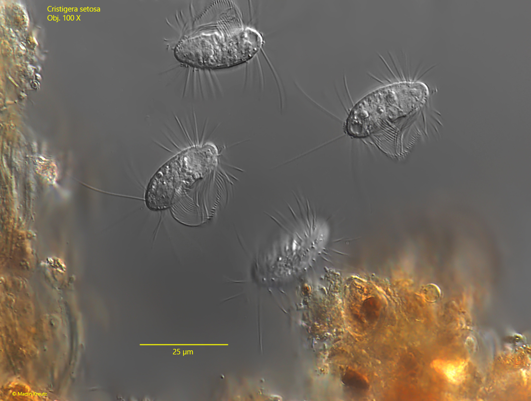
Fig. 1:Cristigera setosa. L = 24–26 µm. A group of of freely swimming specimens between detritus flakes. Obj. 100 X.
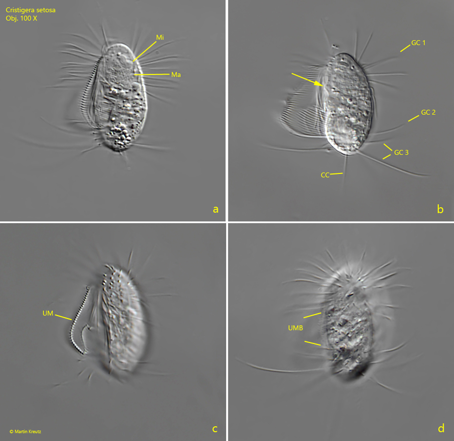
Fig. 2 a-d:Cristigera setosa. L = 28 µm. Different focal planes of a specimen from ventral. Note the cilia grouped in three girdles (GC 1-3). The middle girdle (GC 2) is a diagonal row of single cilia. The cilia of the posterior girdles GC 2 and GC 3 are curved anteriorly. A tooth-shaped projection is found on the right edge of the peristome (arrow, b). CC = caudal cilium, Ma = macronucleus, Mi = micronucleus, UM = undulating membrane, UMB = unciliated mid-body. Obj. 100 X
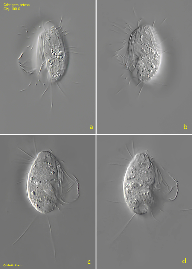
Fig. 3 a-d:Cristigera setosa. L = 28 µm. A second specimen from ventral (a, b) and from dorsal (c, d). Obj. 100 X
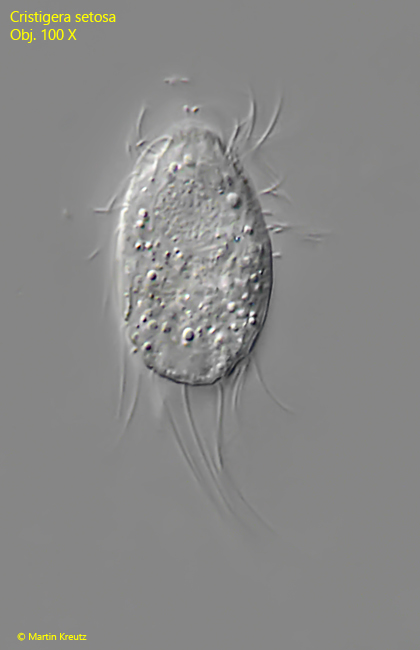
Fig. 4 a-c:Cristigera setosa. L = 27 µm. A third freely swimming specimen from ventral. Note the unciliated mid-body (UMB) between the anterior and posterior ciliation. CC = caudal cilium, CV = contractile vacuole, Ma = macronucleus, Mi = micronucleus, UM = undulating membranelle. Obj. 100 X
Fig. 5:Cristigera setosa. L = 30 µm. A third specimen from ventral. CC = caudal cilium, CV = contractile vacuole, FP = frontal plate, GC 1- 3 = girdles of cilia, Ma = macronucleus. Obj. 100 X
Fig. 6:Cristigera setosa. L = 28 µm. When swimming, the cilia of the posterior girdles are folded into a tail. Obj. 100 X