cells ovoid and rigid, dorso-ventrally slightly flattened
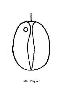
broad side with a longitudinal sulcus (like a coffee bean)
length about 13 µm, width about 10 µm
one or two chloroplasts without pyrenoid
shield-shaped paramylon grains located laterally
one long flagellum
one red eyespot
Cryptoglena australis
I have found Cryptoglena australis in Simmelried only three times so far. First in December 2007, then in November 2019, and most recently in October 2021. This small euglenid flagellate has a longitudinal sulcus on its frontal side, so its assignment to Cryptoglena is clear. The species of the genus described so far are inadequately defined, in my opinion, because it is not clear to what extent there is overlap due to variations in body shape. The specimens I examined were all flattened, making them narrower in lateral view than in frontal view (s. figs. 1d and 2b). This feature best fits Cryptoglena australis, although this species has only been found in Australia so far. The best described species of this genus is Cryptoglena pigra, which is said to possess a conically tapering posterior end. However, I could not clearly identify this feature in my population. Also, Cryptoglena pigra is not supposed to be flattened like my specimens.
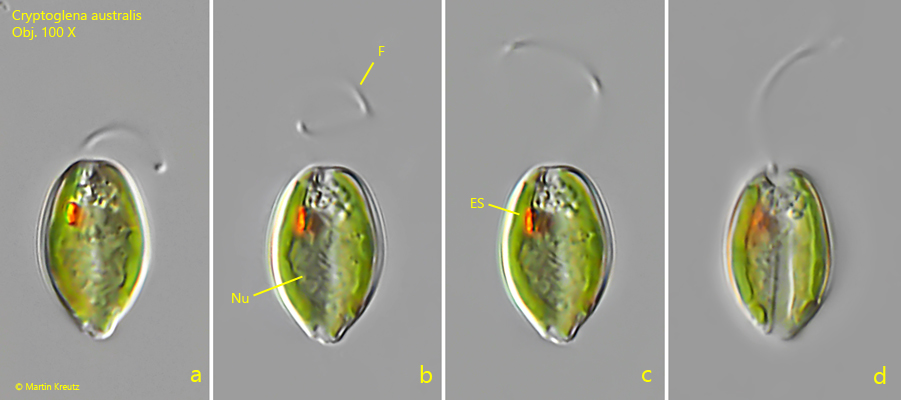
Fig. 1 a-d:Cryptoglena australis. L = 15 µm. Different focal planes of a freely swimming specimen from dorsal. ES = eyespot, F = flagellum, Nu = nucleus. Obj. 100 X.
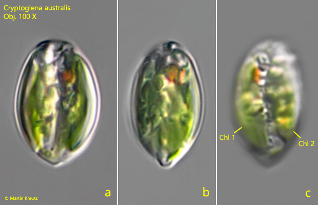
Fig. 2 a-c:Cryptoglena australis. L = 17 µm. Frontal (a), lateral (b) and dorsal view (c) of a second specimen. The dorsal view proves that two chloroplasts are present (Chl 1, Chl 2), as a clear gap is visible between them. Obj. 100 X.
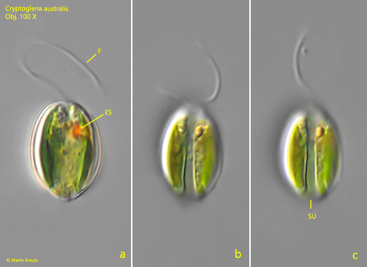
Fig. 3 a-c:Cryptoglena australis. L = 15 µm. Different focal planes of the frontal view of a third, freely swimming specimen. Note the longitudinal sulcus (SU). ES = eyespot, F = flagellum. Obj. 100 X.