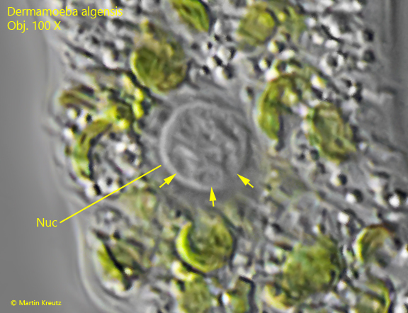
one nucleus (2.9–5.7 µm) with a large, spherical nucleolus (1.4–4.6 µm)
nucleolus with several lacunae
one contractile vacuole, terminally locted
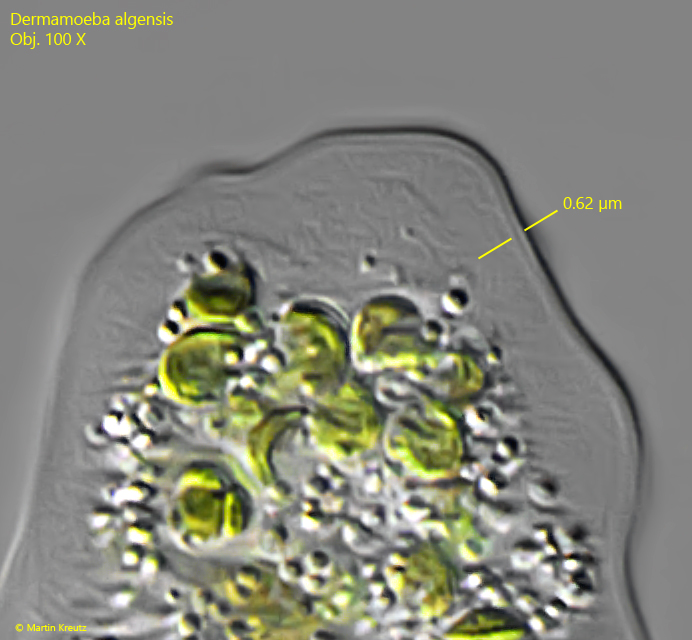
cell coat of 0.5 µm thickness
inconspicuous, bulbous uroid
No drawings from previous authors available.
I have only found a single specimen of Dermamoeba algensis in January 2008. It was found in the top layer of mud in the Simmelried. The only other known locality of Dermamoeba algensis is a small pond near the village of Kirovsk in Russia, where Gromov found the amoeba for the first time in 1999.
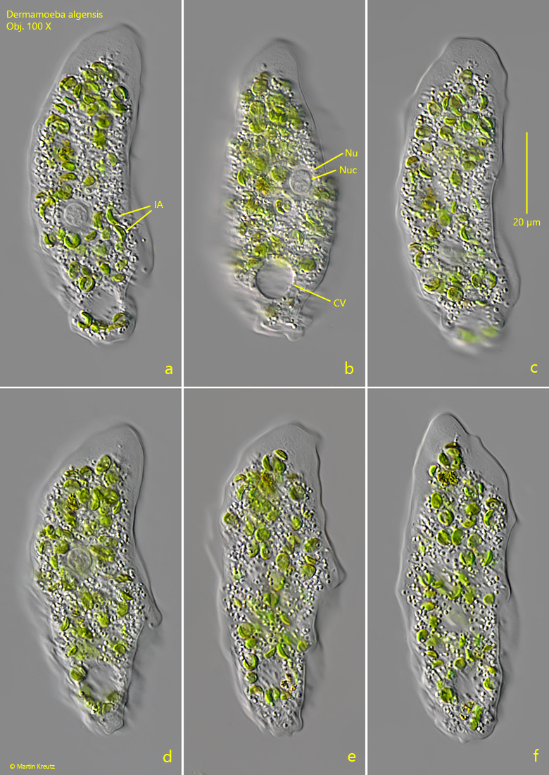
The specimen moved exclusively monopodially with few longitudinal folds on the surface (s. fig. 1 a-f). At first I thought the amoeba was a Thecamoeba. On closer inspection, however, a clear cell coat was visible (s. fig. 2) and is consistent with the description of Dermamoeba algensis by Smirnov et al. (2011). Smirnov et al. summarized all thecamoebic amoebae with a cell coat in the genus Dermamoeba.
My specimen of Dermamoeba algensis was 82 µm long and there were many food vacuoles with algae in the cytoplasm. The nucleus had a diameter of 8.4 µm and was located approximately in mid-body. In the center of the nucleus was a spherical nucleolus with a diameter of 6 µm. It had typical lacunae, as described by Smirnov et al. (s. fig. 3). The nucleus as well as the nucleolus were slightly larger than described by Smirnov et al. but the other features described were the same. The contractile vacuole was located at the posterior end. A poorly defined uroid was formed during locomotion. The amoeba had a clear cell coat of 0.62 µm thickness (s. fig. 2).
Fig. 1 a-f:Dermamoeba algensis. L = 82 µm. Different stages of locomotion. CV = contractile vacuole, IA = ingested algae, Nu = nucleus, Nuc = nucleolus. Obj. 100 X
Fig. 2:Dermamoeba algensis. The cell coat of the specimen shown as in fig. 1 a-f has a thickness of 0.62 µm. Obj. 100 X
Fig. 3:Dermamoeba algensis. The nucleolus (Nuc) with several lacunae (arrows). Obj. 100 X