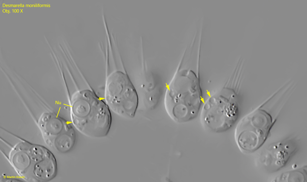
nucleus near anterior end with a central nucleolus
2 contractile vacuoles posteriorly located
Desmarella monoliformis
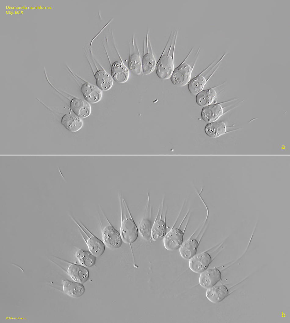
So far I have only found Desmarella moniliformis in the Purren pond and the Simmelried. The colonies gather in large numbers on the surface of the samples. In most cases the colonies are clearly curved. I was also able to find spherical colonies in which the cells were connected in the center. However, these colonies, consisting of a maximum of 16 cells, disintegrated under the coverslip.
Most colonies consisted of 8–16 cells. Colonies with fewer cells (sometimes only 2) were also present. The individual cells were 12–13 µm long and thus considerably larger than described for Desmarella moniliformis (about 6 µm). With this size they would rather correspond to Desmarella brachycalyx (9.5–11 µm), but this species is said to form colonies of only 4 cells. Only for Desmarella moniliformis colonies of the size I have found are described. I think it is also possible that Desmarella moniliformis and Desmarella brachycalyx are synonymous, because colony size and body length of the individual cells can be subject to great variability.
The nucleus of Desmarella moniliformis is located at the anterior end and has a clear, central nucleolus and a thick nuclear membrane (s. figs. 2 a and 3). The contractile vacuoles are not easy to recognize and in my population they were not located at the posterior end but at the cell equator (s. fig. 3). I could find a maximum of 2 contractile vacuoles per cell, as described for Desmarella moniliformis. At the posterior end of the cells usually large food vacuoles are located (s. fig. 2 a). These may have been mistaken for the contractile vacuoles by earlier authors. In my population the collar of parallel microvilli was 8.5 – 10.5 µm high (s. fig. 2 b).
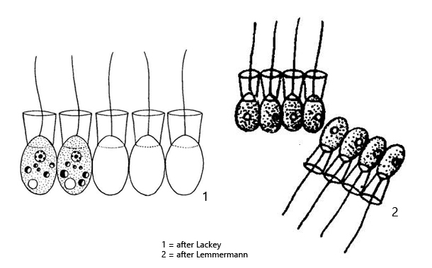
All cells of the colonies I observed were radially connected with thin stalks at a central starting point (s. fig. 2 a). To my knowledge, these stalks have not yet been described. They were also not shown in drawings of Desmarella moniliformis by earlier authors (s. drawings above).
Fig. 1 a-b:Desmarella moniliformis. L = 12–13 µm (of cells). Two focal planes of a slightly squashed colony of 13 cells. Obj. 60 X.
Fig. 2 a-b:Desmarella moniliformis. L = 12–13 µm (of cells). The colony as shown in fig. 1 a-b in detail. The cells are connected radially with delicate stalks (ST, only three in focal plane). The large food vacuoles (FV) are located mainly posterior. The base of the flagella (F) is surrounded by a circular collar (CO) of microvilli with a height of 8.5–10.5 µm. NU = nucleus. Obj. 100 X.
Fig. 3:Desmarella moniliformis. L = 12–13 µm (of cells). A part of the colony as shown in fig. 2 a-b with focal plane on the contractile vacuoles (arrows). Note the central nucleolus in each nucleus (Nu). Obj. 100 X.