linear coenobia of 2–8 cells, arranged tightly in parallel cells 10–36 µm long, width 3–8 µm
cells cylindrically shaped with rounded ends
inner and outer cells about equal in length
poles of the outer cells with 4 long spines (8–18 µm)
sometimes small warts arranged diagonally at the poles of the inner cells
one pyrenoid per cell
Desmodesmus communis
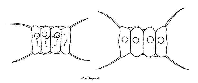
Desmodesmus communis was first described by Turpin (1820) as Scenedesmus quadradricauda. Hegewald (1977) recognized that the species Scenedesmus quadricauda was not sufficiently described and classified by Turpin, so he proposed the new name Scenedesmus communis. However, in 2000 the species was transferred to the new genus Desmodesmus by Hegewald based on DNA analysis.
I find Desmodesmus communis very frequently and regularly in the plankton of the pond of the waste disposal company of Constance. The species can the identfied by the evenly spaced cells that are all about the same length. This results in an almost rectangular shape of the coenobium. The incisions between the cells are not deep. The 4 spines on the outer cells are about as long as the cells and sometimes brownish in color (s. fig. 1). At the poles of the middle cells, small warts are sometimes found, arranged diagonally (s. fig. 1). In addition, the inner cells are often surrounded by a tightly fitting mucilaginous sheath (s. fig. 2 a).
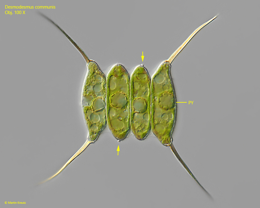
Fig. 1:Desmodesmus communis. L = 38 µm (of coenobium). A slightly squashed coenobium of 4 cells. The cells are 28 – 30 µm long and the 4 spines are 27 – 29 µm long. Note the diagonally arranged warts at the poles of the inner cells (arrows). Obj. 100 X.
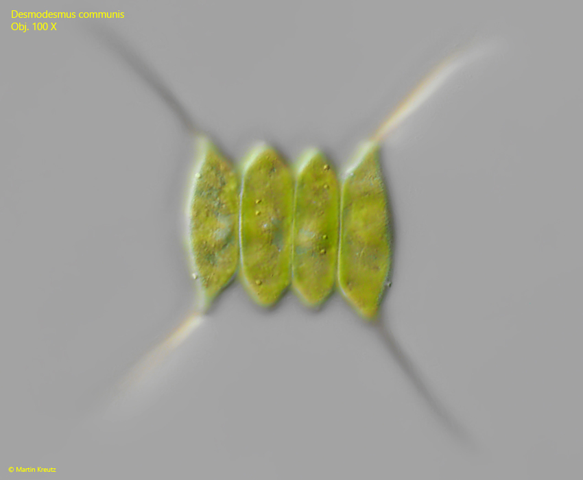
Fig. 2:Desmodesmus communis. L = 38 µm (of coenobium). The same specimen as shown in fig. 1 with focal plane on the cells surface. The cells surface is smooth apart from some small warts. Obj. 100 X.
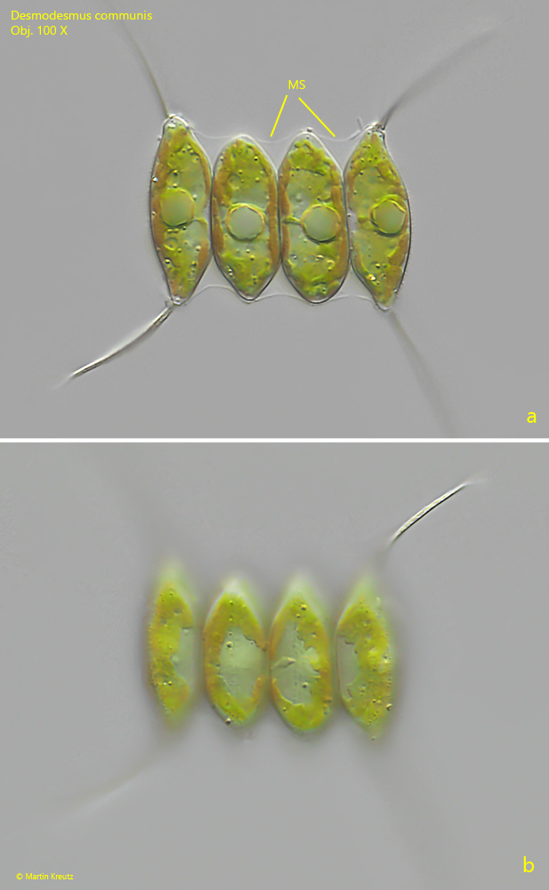
Fig. 3:Desmodesmus communis. L = 36 µm (of coenobium). Two focal planes of a second specimen. The cells are 24 – 26 µm long. Note the mucilage sheath (MS) covering the inner cells. Obj. 100 X.