linear coenobia of 4–8 cells, arranged tightly in parallel cells 12–34 long, width 4–7 µm
cells cylindrically shaped with rounded ends
inner cells elongated oval or spindle-shaped, shorter than outer cells
outer cells oval in middle with capitate apices
inner cells with with rounded apices, often with small spines
distinct main spines at apices of outer cells straight or sigmoidal curved
one pyrenoid per cell
Desmodesmus protuberans
Desmodesmus protuberans was originally described by Fritsch & Rich (1929) as Scenedesmus protuberans but was transferred to the genus Desmodesmus by Hegewald in 2000.
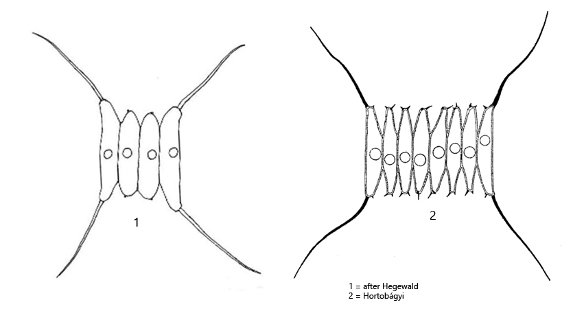
In September 2022 I found Desmodesmus protuberans in the plankton of the pond of the waste disposal company Constance. I found coenobia with 4 but also with 8 cells. The outer cells are slightly longer than the inner cells and the apices of them are characteristically slightly bent outwards and somewhat thickened. In addition, the cell ends of the outer cells bear very long, often brownish or black-colored spines, which can become as long as the coenobium. The inner cells often have small spines at their ends, but can also be naked. In my population, all the specimens I examined had spines on the apices of the inner cells. In addition, in some specimens I was able to observe weakly developed ribs on the sides of the inner cells, which reached about a quarter of the cell length (s. fig. 1 b). To my knowledge, these ribs have not been described by earlier authors.
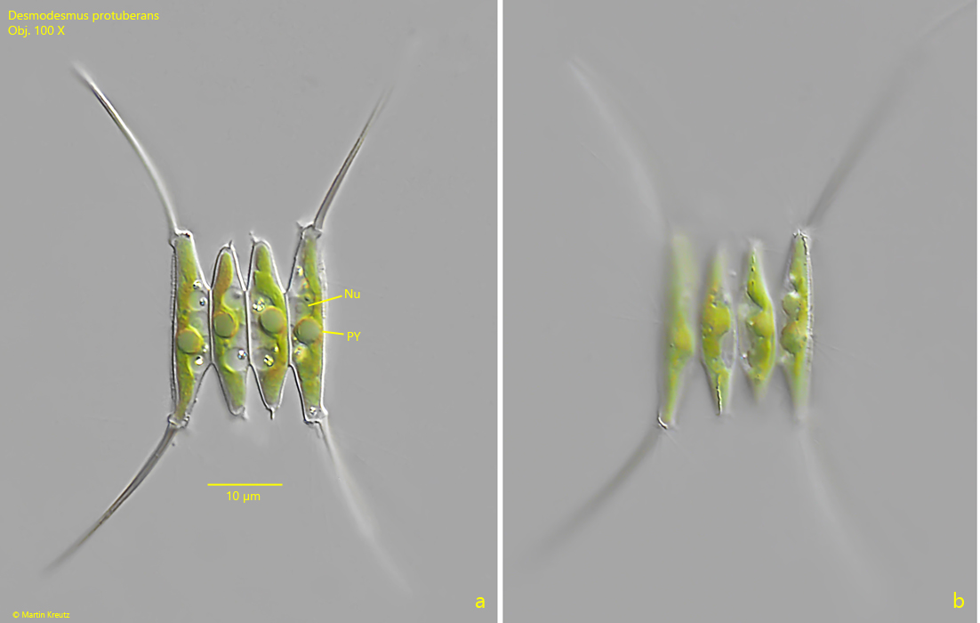
Fig. 1 a-b:Desmodesmus protuberans. L = 20 µm (of coenobium). Two focal planes of a coenobium of 4 cells. Nu = nucleus, PY = pyrenoid. Obj. 100 X.
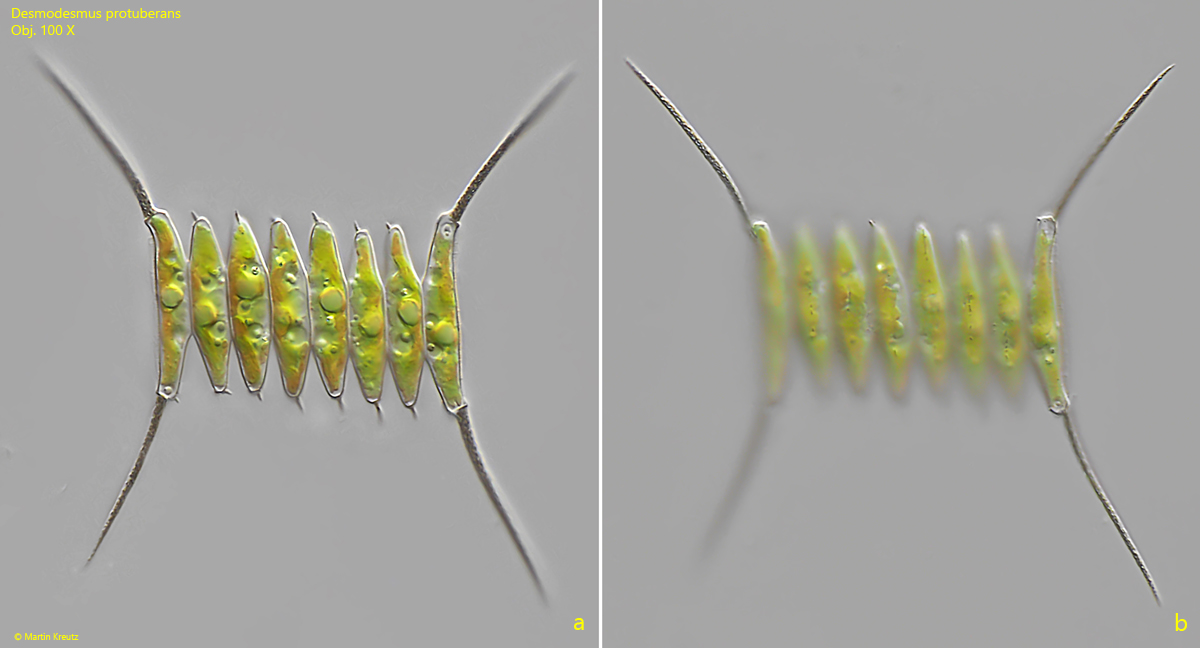
Fig. 2 a-b:Desmodesmus protuberans. L = 43 µm (of coenobium). Two focal planes of a coenobium of 8 cells. Obj. 100 X.