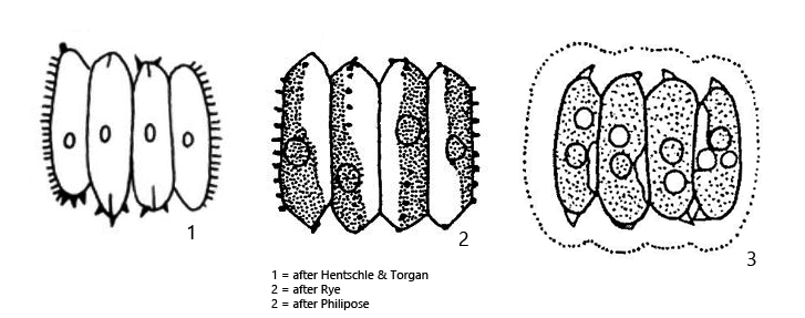
often a row of small teeth along the lateral wall of the outer cells
cell wall smooth (apart from teeth)
one pyrenoid per cell
Desmodesmus serratus
Desmodesmus serratus was originally described by Bohlin (1901) as Scenedesmus serratus, but was transferred to the genus Desmodesmus by An, Friedl and Hegewald in 1999.
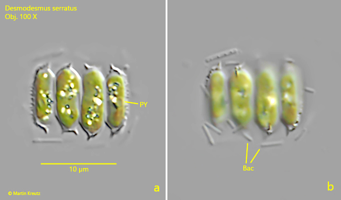
In August 2023 I found Desmodesmus serratus in large numbers in the plankton of the pond of the waste disposal company Constance. This species is very small and can easily be overlooked when sifting through a plankton sample. Desmodesmus serratus can be recognized by the very small teeth on the apices of the cells. In addition, a mucus sheath around the coenobium is characteristic, which only becomes visible in DIC by attached bacteria (s. fig. 2 a-b). A row of small teeth can often be found on the lateral outer sides of the coenobium (s. fig. 1 b). Otherwise the walls of the cells are naked (s. fig. 2 b). The similar species Desmodesmus brasiliensis has longitudinal ridges on the lateral sides of the cells in addition to the apical, small teeth. This allows to distinguish these two species.
Fig. 1 a-b:Desmodesmus serratus. L = 11.6 µm (of coenobium). Two focal planes of a coenobium of 4 cells. Obj. 100 X.
Fig. 2 a-b:Desmodesmus serratus. L = 12.6 µm (of coenobium). Two focal planes of a second coenobium of 4 cells. Note the bacteria (Bac) covering the mucilage sheath of the coenobium. PY = pyrenoid. Obj. 100 X.