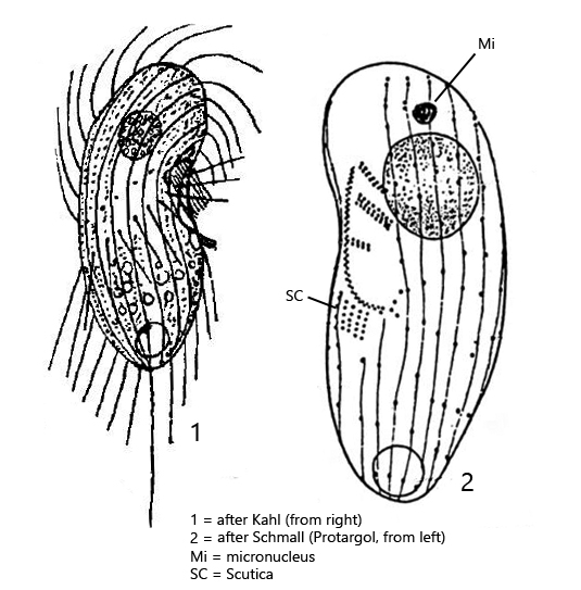
length 30–45 µm macronucleus spherical in anterior half
one spherical micronucleus in anterior fifth of cell
contractile vacuole terminal
cytoplasm often filled with symbiotic bacteria
sometimes extrusomes present
about 20 longitudinal rows of cilia
frontal plate and praeoral area without cilia
a cilia-free zone in mid-body
one caudal cilium of body legth
oral apparatus large, in anterior third shifted to the left side
below oral apparatus a field of basal bodies, presumably the scutica
Dexiotrichides centralis
By earlier authors Dexiotrichides centralis is described as a very common species, especially in nutrient-rich waters. However, this ciliate was described by Foissner, Berger and Kohmann in 1994 in their “Revision der Ciliaten des Saprobiensystems, Band II” (s. literature) and it was stated: “wir haben diese Art noch nie gesehen! (However, we have never seen this species!)”. In fact, I have also been able to find only 4 specimens of Dexiotrichides centralis in my sampling sites within 25 years. It remains unclear whether the species has become generally rarer in the last 100 years or whether the wrong finding areas were sampled.
Based on these 4 specimens I could document the main characteristics of Dexiotrichides centralis. In my population, all specimens had a very long caudal cilium (about body length, s. fig. 1a). In addition, all specimens contained very large, 7–8 µm long, symbiotic bacteria (s. fig. 3b and 4). They must be symbiotic bacteria, because I could find food vacuoles co-occuring with these bacteria in the cytoplasm (s. fig. 4). Thus, the 7–8 µm long bacteria were not ingested as food. As described by Schmaller from protargol preparations (s. drawing above), the micronucleus is located in the anterior fifth of the cell and is hard to find (s. fig. 5). On the left side a small field of basal bodies without cilia is located below the mouth opening. This is probably the so-called scutica (s fig. 3a). From this field of basal bodies the oral apparatuses of the two daughter cells are organized during cell division. On the right side of the body I could observe an oblique cilia-free zone as drawn by Kahl (s. drawing above and fig. 6).
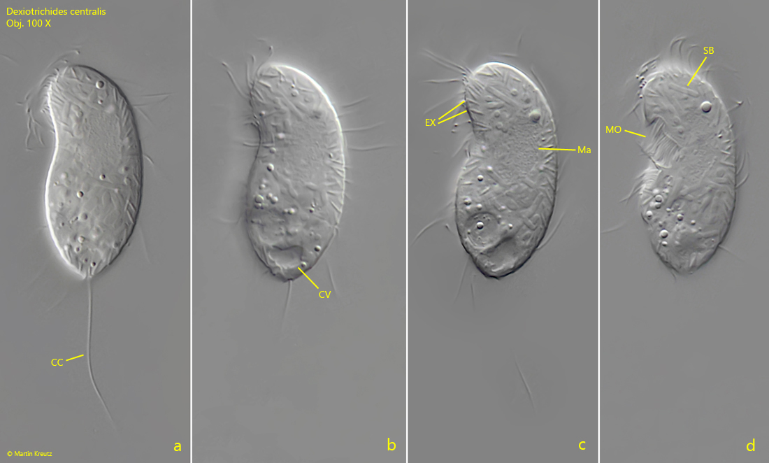
Fig. 1 a-d:Dexiotrichides centralis. L = 38 µm. Different focal planes of a freely swimming specimen from left. Obj. 100 X.
Fig. 2 a-b:Dexiotrichides centralis. L = 44 µm. Two focal planes of a freely swimming specimen from ventral. Note the mouth opening (MO) shifted to the left side. Obj. 100 X.
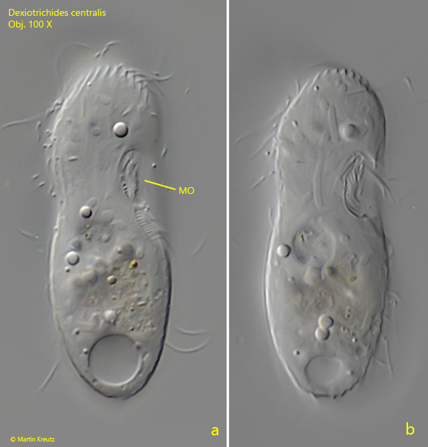
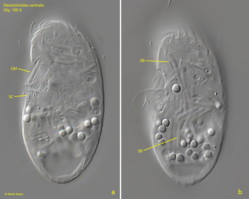
Fig. 3 a-b:Dexiotrichides centralis. L = 42 µm. Two focal planes of a slightly squashed specimen from the left side. Note the cilia-free field of basal bodies below the mouth opening, presumably the so called scutica (SC). The body is filled with large, about 7–8 µm long, symbiotic bacteria (SB). UM = undulating membrane. Obj. 100 X.
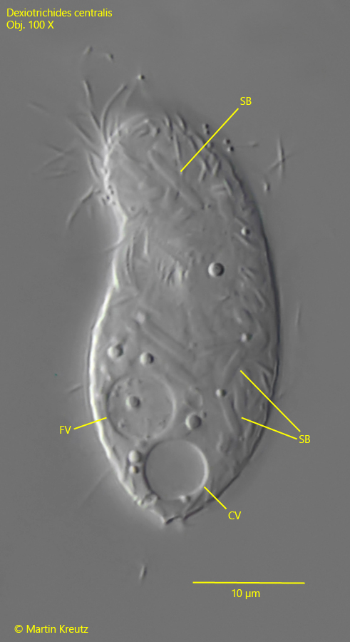
Fig. 4:Dexiotrichides centralis. L = 38 µm. The specimen shown in fig. 1 a-d with focal plane on the symbiotic bacteris (SB) scattered in the cytoplasm. The co-occuring of a food vacuole (FV) proves, that the large bacteria in the cytoplasm are not ingested as food. Obj. 100 X.
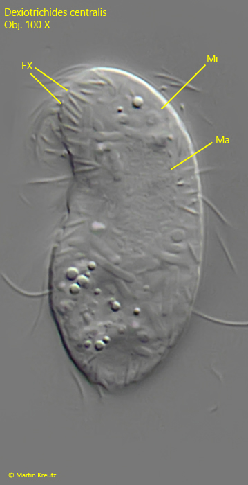
Fig. 5:Dexiotrichides centralis. L = 38 µm. The specimen shown in fig. 1 a-d with focal plane on the micronucleus (Mi) in the anterior fifth of the body. Without staining it is hard to see. This specimen has a fringe of 3 µm long extrusomes (EX). Ma = macronucleus. Obj. 100 X.
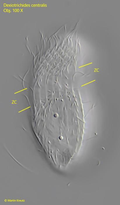
Fig. 6:Dexiotrichides centralis. L = 45 µm. A slightly squashed specimen with focal plane on the ciliation of the right side. Note the cilia-free zone (ZC) in the mid-body region. Obj. 100 X.