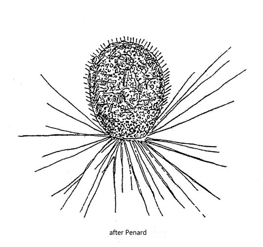
test ovoid or pear-shaped, membranous and flexible, brownish color
test covered with organic and inorganic material
hyaline, transparent rods adhere to the test, 8–10 µm long
length 25–178 µm
aperture of test broad, circular or oval
nucleus in posterior end of the test
one or thwo contractile vacuoles
filiopodia branching and anstomosing, arising in all directions
Diaphoropodon mobile
I have found Diaphoropodon mobile so far only in the Sima Moor (Austria) in the Schwemm (Moor, Austria) and in the Simmelried. However, in Simmelried I have not been able to detect the species in the last 7 years.
The most conspicuous feature of Diaphoropodon mobile are thin, hyaline rods with which the test is covered. The purpose of these rods is unknown. They are absent in empty tests of dead specimens. Thus, it is possible that these rods are formed by the amoeba and protrude through the test. The length of the rods is reported to be 8-10 µm. However, in the specimens I examined, the length varied considerably between 4–18 µm (s. figs. 1, 2 and 3 a-b). If the rods are really different in length or if they protrude through the test with different lengths due to different thicknesses of the test cannot be determined.
In fresh samples the specimens are difficult to find, because they are often masked by detritus. In my experience it is best to create microaquaria with high layer thickness and fill them with detritus containing samples. After about 1–2 days specimens can be found outside the detritus. Either isolate these specimens or try to reduce the layer thickness of the microquariums as far as the foreign bodies under the coverslip allow.
The division of Diaphoropodon mobile ocurrs along the longitudinal axis (s. fig. 4). The division begins at the posterior end. Not only does the protoplast divide, but the flexible test constricts and also divides.
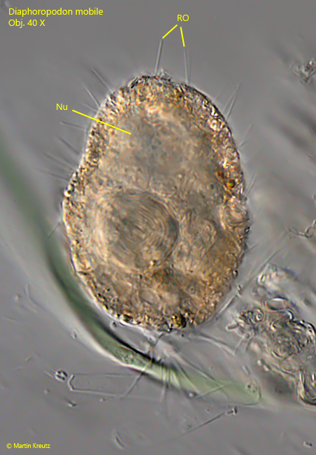
Fig. 1:Diaphoropodon mobile. L = 124 µm. A specimen with extended filopodia and 16–18 µm long rods (RO). Nu = nucleus. Obj. 40 X.
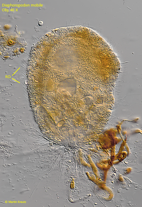
Fig. 2:Diaphoropodon mobile. L = 138 µm. The test of this specimen is mainly covered with organic material. The rods (RO) are 9–10 µm long. Obj. 40 X.
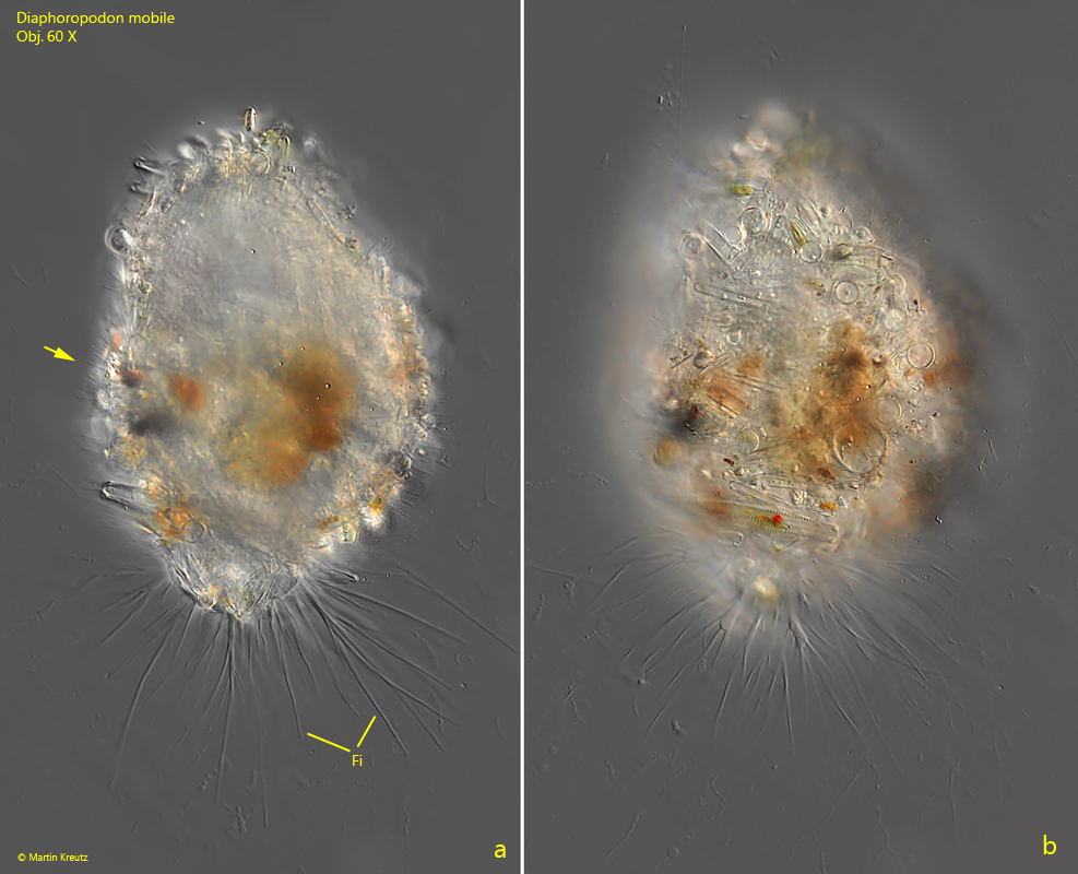
Fig. 3 a-b:Diaphoropodon mobile. L = 145 µm. Two focal planes of a specimen with extended and branched filopodia (Fi). The rods (arrow) of this specimen are only 4–5 µm long. Obj. 60 X.
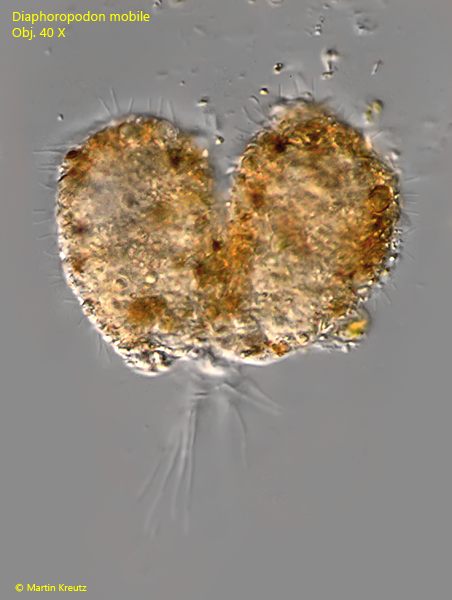
Fig. 4:Diaphoropodon mobile. L = 65 µm. A specimen during cell division along the longitudinal axis. Obj. 40 X.