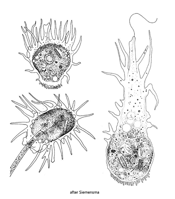
cells ovoid or pyriform in locomotion, slow movement
resting cells spherical
numerous conical pseudopodia
forms in locomotion up to 450 µm long
resting forms about 100–200 µm in diameter
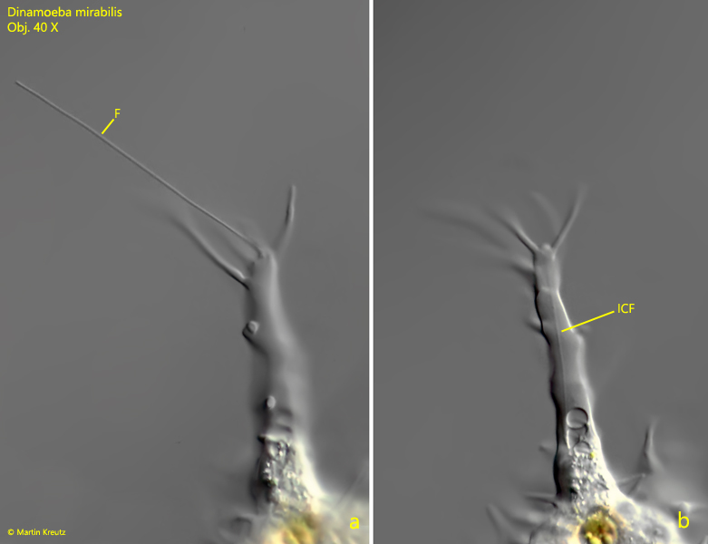
sometimes a flagellum is present with a continuation inside the plasm
ectoplasm clear and hyaline
large spherical nucleus 25–40 µm in diameter
nucleus surrounded by a thick layer of symbiotic bacteria
feeds on algae and detritus
a few contractile vacuoles
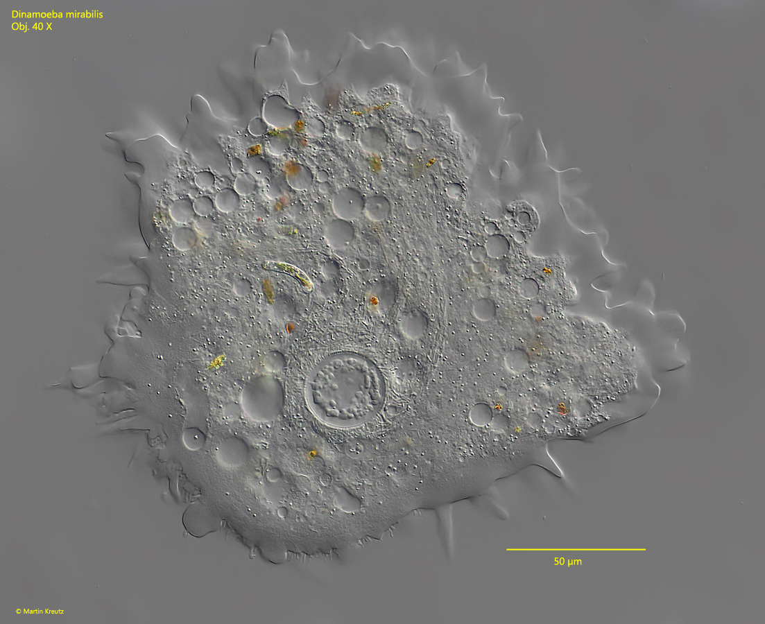
Dinamoeba mirabilis
I have found Dinamoeba mirabilis only in Simmelried, where it is regularly found in mud and decomposing plant masses. The sizeable amoeba is easily recognized by a thick layer of symbiotic bacteria around the nucleus (s fig. 3). Except during division processes (s. figs. 4-6) it has only one nucleus, which is always filled with cloudy nucleoli. In the literature this species is also described with 2 nuclei, but I have never found this in my population. Another characteristic feature is the flagellum, which has a clear continuation in the plasm (s. fig. 9). The flagellum is mostly stretched out straight and periodically performs whip-like movements. Because of the flagellum Dinamoeba mirabilis is included in the Mastigamoebidae, but the flagellum is not always present. Often resting specimens can be found without a flagellum. In addition to the large, rod-shaped bacteria around the nucleus, many thinner, rod-shaped bacteria are found in the plasm (s. fig. 3). This composition of symbiotic bacteria is reminiscent of Pelomyxa. According to Leidy, the cell surface of Dinamoeba mirabilis is supposed to be covered with bacteria-like rods. I could not observe this in my population. All specimens I have observed had a smooth surface.
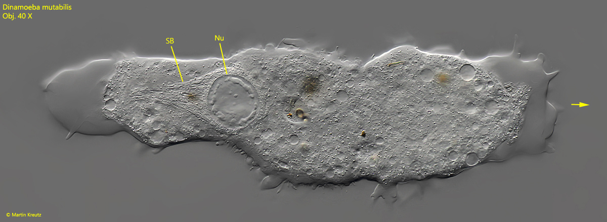
Fig. 1: Dinamoeba mirabilis. L = 450 µm. A specimen during locomotion in limax form. arrow = locomotion dirction, Nu = nucleus, SB = layer of symbiotic bacteria surrounding the nucleus. Obj. 40 X.
Fig. 2:Dinamoeba mirabilis. D = 250 µm. A resting specimen. Obj. 40 X.
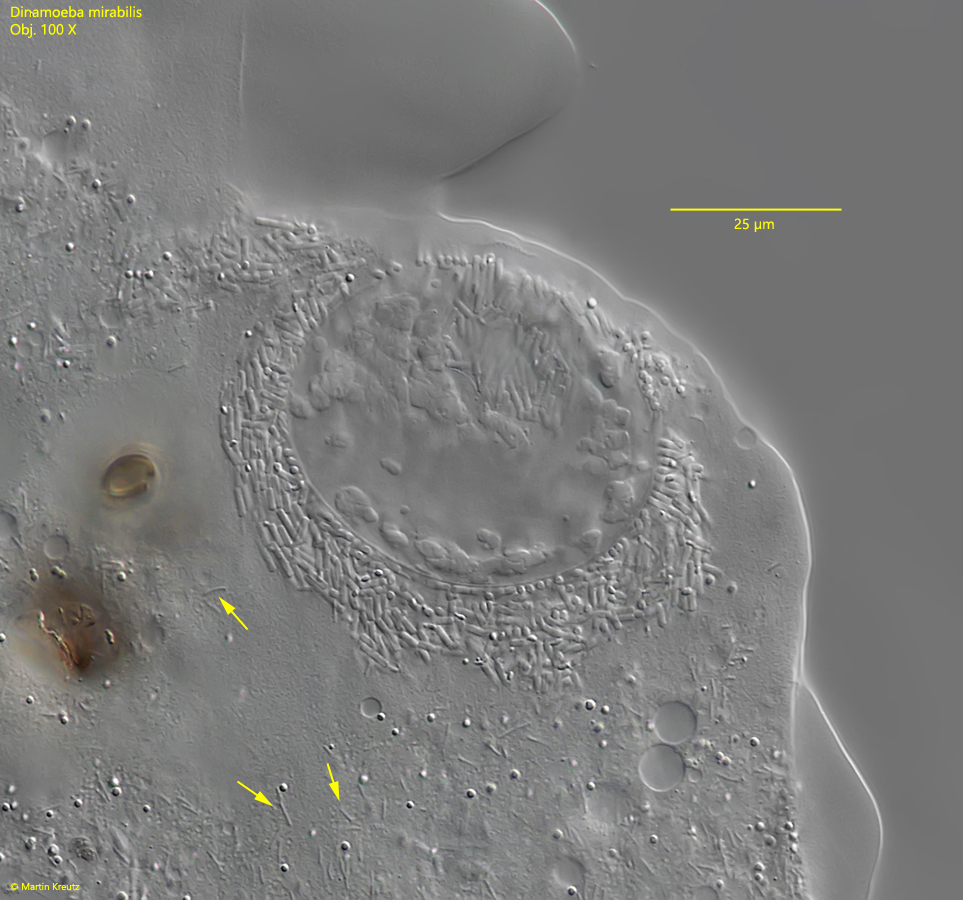
Fig. 3: Dinamoeba mirabilis. The nucleus is covered with a thick layer of rod shaped symbiotic bacteria. The length of these bacteria is 4-6- µm. Note the cloudy nucleoli in the nucleus and the thin rods of symbiotic bacteria scattered in the plasm (arrows). These thin symbiotic bacteria are 3 – 4 µm long. Obj. 100 X.
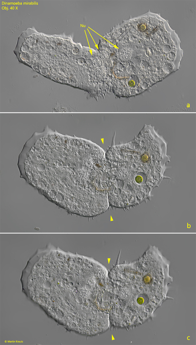
Fig. 4 a-c: Dinamoeba mirabilis. L = 240 µm. A specimen with 4 nuclei in the process of cell division. Arrowheads = dividing furrow, Nu = nuclei. Obj. 40 X.
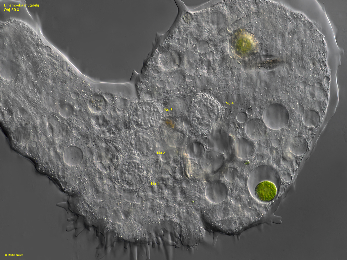
Fig. 5: Dinamoeba mirabilis. L = 240 µm. A closer view of the 4 nuclei in the dividing specimen shown in fig. 4 a-c. Obj. 60 X.
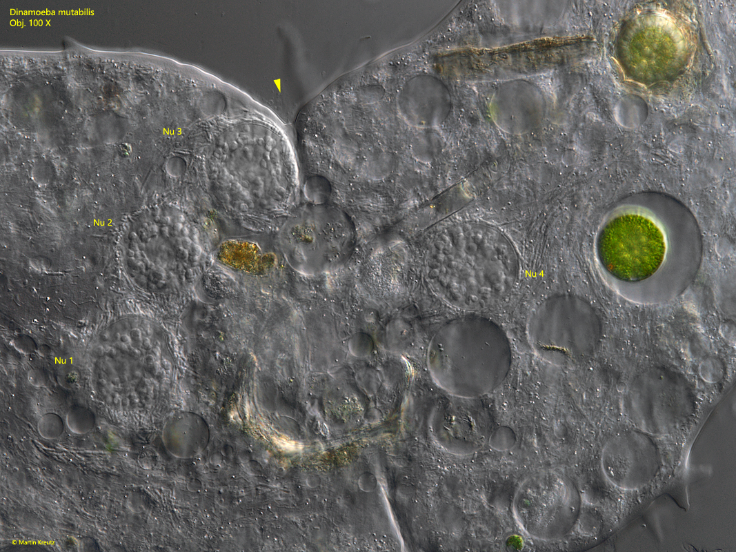
Fig. 6: Dinamoeba mirabilis. L = 240 µm. The 4 nuclei of the specimen in division in detail. The division furrow (arrowhead) is clearly visible. Obj. 100 X.
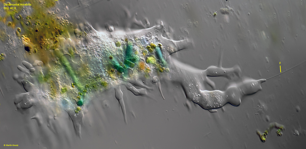
Fig. 7: Dinamoeba mirabilis. L = 320 µm. A freely floating specimen with a flagellum (F). The flagellum arises from the tip of a pseudopodium, which is extended in the direction of flow. Obj. 40 X.
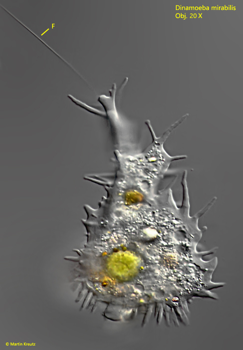
Fig. 8: Dinamoeba mirabilis. L = 300 µm. A second freely floating specimen with a flagellum (F). Note the pyriform shape of the specimen. Obj. 20 X.
Fig. 9 a-b: Dinamoeba mirabilis. L = 300 µm. The straight flagellum (F) is 190 µm long and has a continuation within the plasm of the pseudopodium (ICF). Obj. 40 X.