cells attached with a tapered stalk of cytoplasm to the posterior part of the lorica
two golden brown chloroplasts
the anterior chloroplast with an eyespot
two flagella of different lengths
one spherical nucleus between chloroplasts
one contractile vacuoles in midbody
length of lorica 30–40 µm
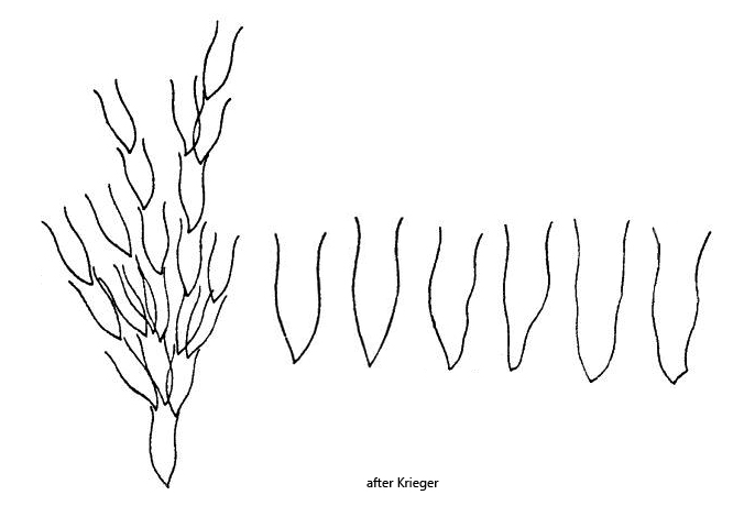
lorica vase-shaped, somwhat bulbous in the middle
cells forming dense and bushy colonies
angle between branches of the colony small
Dinobryon sertularia
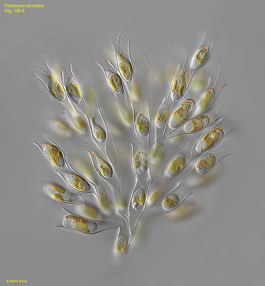
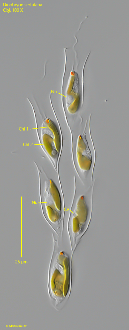
Dinobryon sertularia is by far the most common species of this genus in my sampling sites and is present in almost all plankton samples. Colonies are easily recognized by their dense and bushy shape. The vase-shaped loricae of cellulose are mostly smooth and without wavy undulatuions as in Dinobryon divergens. The middle part of the lorica is bulbous and its opening is slightly flared like a funnel. The cells have two flagella of different lengths and two separate chloroplasts. The anterior chloroplast has an eye spot. In the middle between the chloroplasts is the nucleus. The contractile vacuole is located in the middle of the body.
Fig. 1:Dinobryon sertularia. L = 136 µm (of colony). A slightly squashed colony. Note the bushy shape. Obj. 100 X.
Fig. 2:Dinobryon sertularia. A small colony of 6 cells. Chl 1-2 = chloroplasts, CV = contractile vacuole, Nu = nucleus. Obj. 100 X.
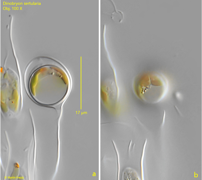
Fig. 3:Dinobryon sertularia. The spherical cyst of a specimen. Obj. 100 X.