body disc-shaped, laterally flattened, with frontal spine
length 70–90 µm
horseshoe-shaped bulge with perizonal stripe in anterior third
perizonal stripe interrupted on left side
adoral zone in a depression below perizonal stripe
two curved spines on right side
one short and curved spine on left side
pellicle rigid, transparent
macronucleus spherical or oval, in posterior third
one spherical micronucleus adjacent to macronucleus
cytoplasm filled with symbiotic bacteria
contractile vacuole in mid-body
ciliation strongly reduced
Discomorphella pectinata
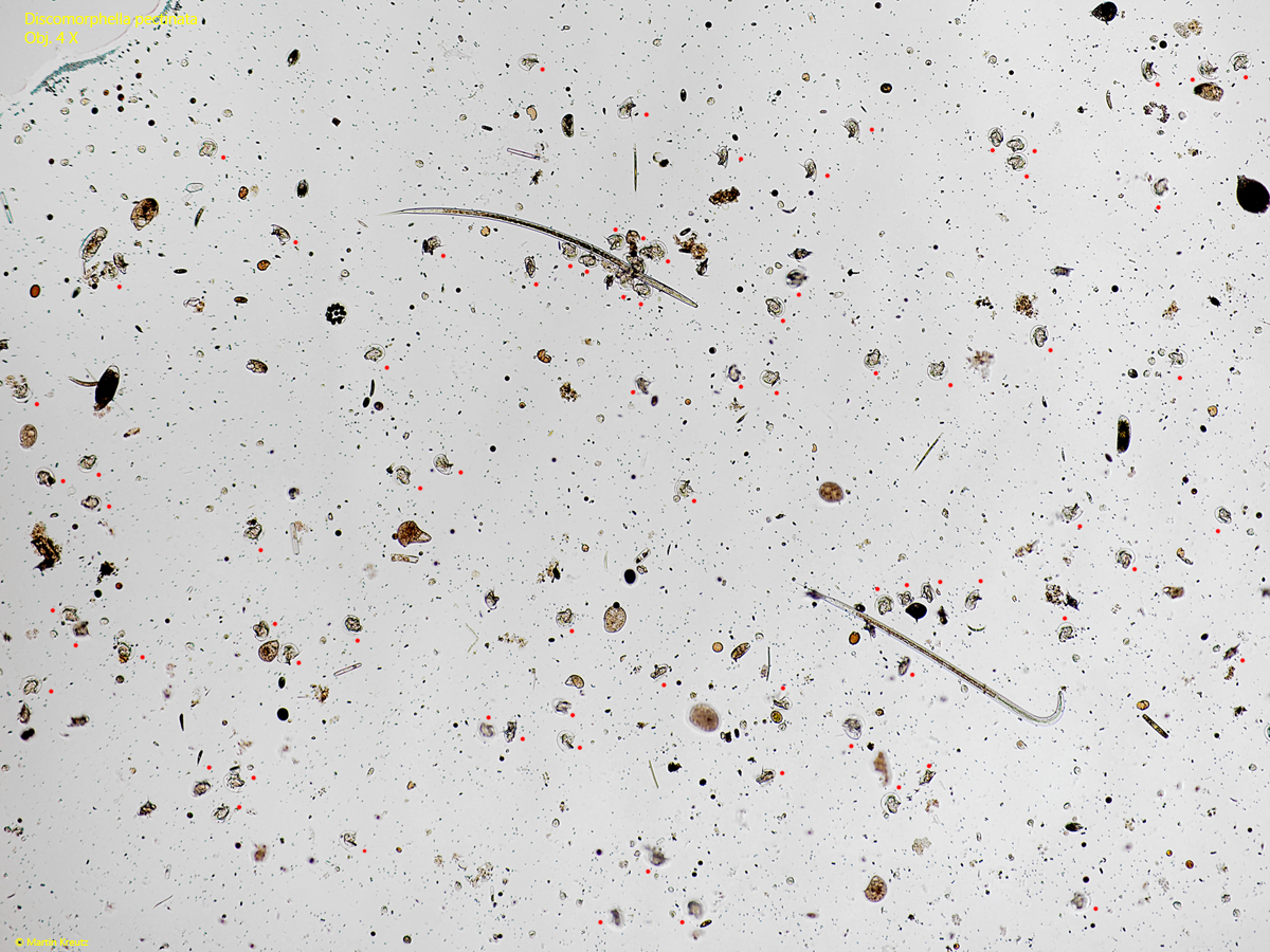
I find Discomorphella pectinata exclusively in the Simmelried, where the species occurs frequently and regularly. In August 2018, there was a mass development of Discomorphella pectinata with a concentration of about 1000 specimens per milliliter (s. fig. 1). Such mass developments did not occur before or after.
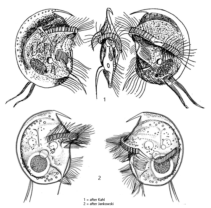
Discomorphella pectinata is unmistakable due to its abstract, asymmetrical shape. In addition to the frontal spine (S1), there are two long, curved spines on the right side (S2, S3) and another short, curved spine (S4) on the left side, which is attached to the separated part of the interrupted perizonal stripe (s. figs. 2 a-c, 3 a-b and 4). The ciliation is reduced to the perizonal stripe, which runs across the ventral side (s. fig. 5 a), a dorsal tuft of cilia and a short ventral row of cilia (s. fig. 6). This makes Discomorphella pectinata a slow and clumsy-looking swimmer. Numerous symbiotic bacteria can be found in the cytoplasm. I was able to identify at least two species (s. fig. 7). Thicker rods with a length of approx. 9–16 µm and thin rods with a length of 6–9 µm. This combination of symbiotic bacteria is also found in other anaerobic ciliates.
Fig. 1:Discomorphella pectinata. Overview of a mass development in August 2018 with a concentration of about 1000 specimens per milliliter. The image shows 78 specimens marked with a red dot. Obj. 4 X.
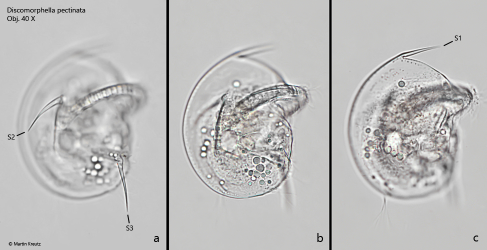
Fig. 2 a-c:Discomorphella pectinata. L = 83 µm. Different focal planes from the right side. Note the frontal spine (S1) and the two lateral spines (S2, S3). Obj. 40 X.
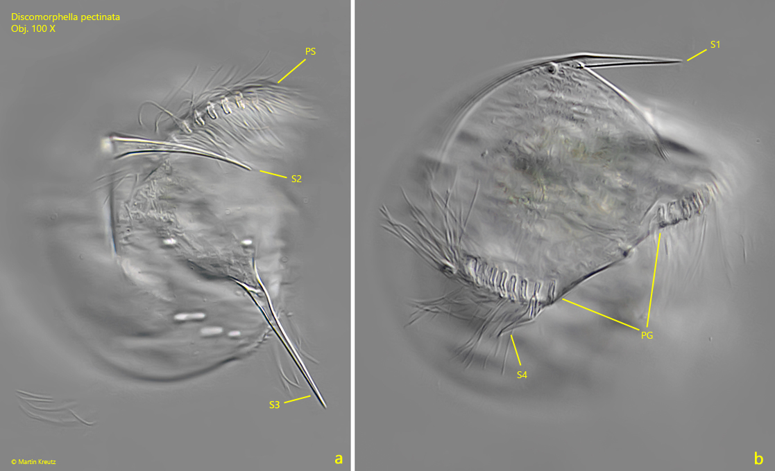
Fig. 3 a-b:Discomorphella pectinata. L = 75 µm. A slightly squashed specimen from the right side (a) and focal plane on the left side (b). Note the perizonal stripe (PS) on a horseshoe-shaped bulge and the gap in the perizonal stripe (PG) on the left side. On the left side is also the short spine S4 visible. Obj. 100 X.
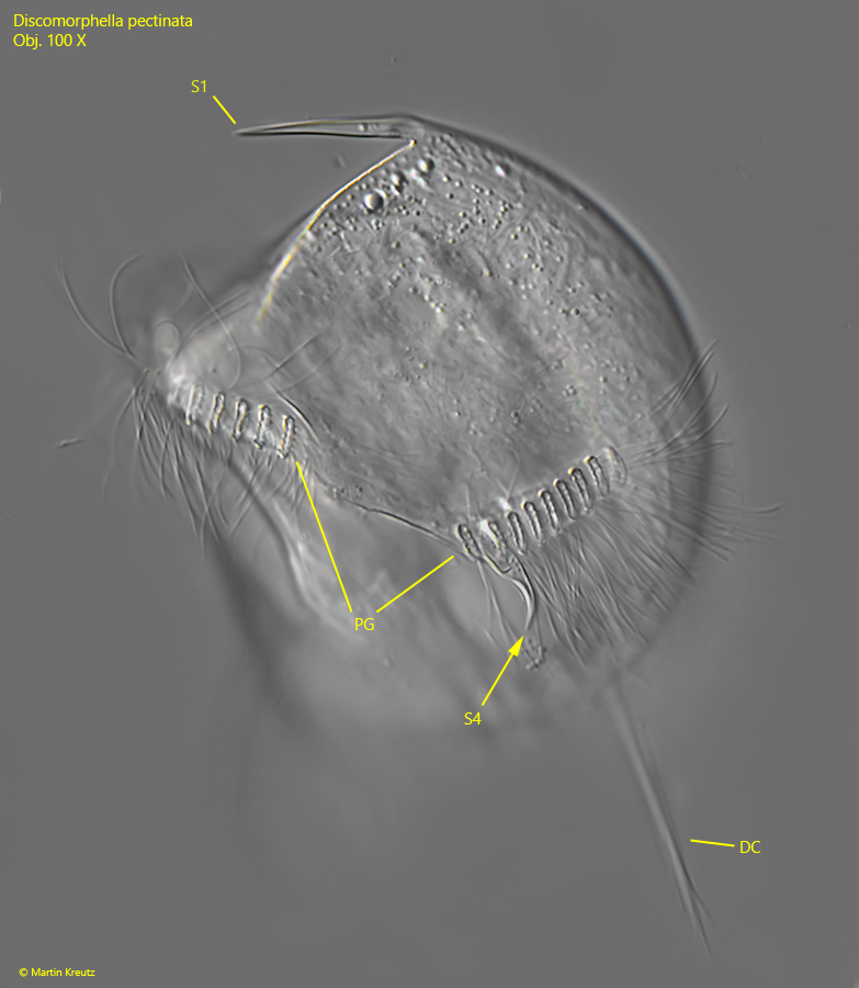
Fig. 4:Discomorphella pectinata. L = 77 µm. A slightly squashed specimen from the left side. DC = tuft of dorsal cilia, PG = gap in the perizonal stripe, S1 = frontal spine, S4 = left spine. Obj. 100 X.
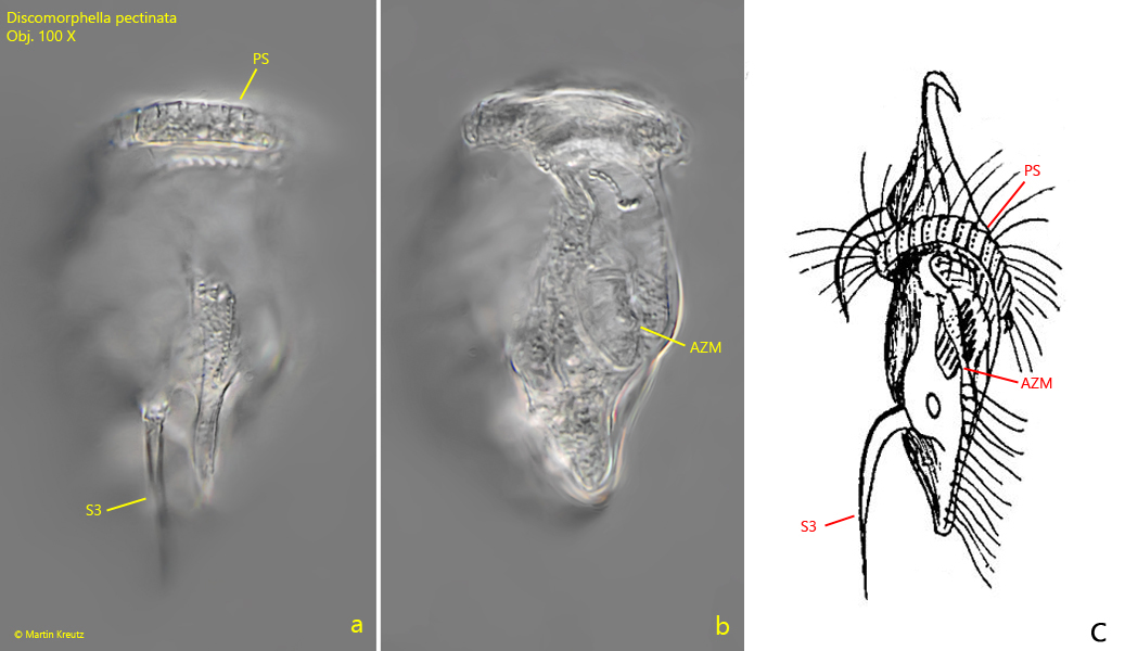
Fig. 5 a-c:Discomorphella pectinata. L = 73 µm. Two focal planes from ventral side (a, b) in comparision with the drawing of Kahl (c). Apically the transverse perizonal stripe (PS) is visible and on the right side the spine 3 (S3). Approximately in the middle of the ventral side lies the adoral zone of membranelles (AZM) in a depression, as drawn by Kahl. Obj. 100 X.
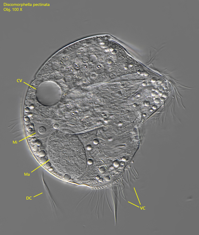
Fig. 6:Discomorphella pectinata. A squashed specimen from right L = 73 µm. The oval macronucleus (Ma) with the adjacent micronucleus (Mi) is visible. CV = contractile vacuole, DC = tuft of dorsal cilia, VC = ventral cilia row. Obj. 100 X.
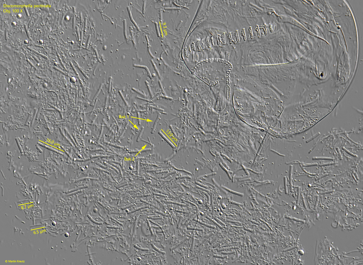
Fig. 6:Discomorphella pectinata. The released symbiotic bacteria of a strongly squashed specimen. The are at least 2 types of symbiotic bacteria (Bak 1, Bak 2). The thick rods of type 1 are 9–16 µm long, while the thin rods of type 2 have a length of 6–9 µm. Obj. 100 X.