shell hyaline, thin, without xenosomes, older cells yellowish or brown
in frontal view spherical or broadly oval, sometimes irregular
in lateral view dome-shaped
shell with two opposite necks
length of shell (including necks) 22–32 µm
length of necks 1.7–3.4 µm
granulofilopodia form a dichotomously branched network
nucleus central, with a single nucleolus, older cells with numerous nucleoli
up to 10 contractile vacuoles
Ditrema longicollis
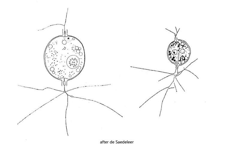
This granulofilose testate amoeba was first described by De Saedeleer as Diplophrys longicollis, but later transferred to Ditrema by Siemensma (s. Ferry Siemensma – Ditrema longicollis). I have found Ditrema longicollis so far exclusively in the Simmelried. It colonizes floating coverslips, where it can be easily observed. The shell with two opposite necks looks spherical or oval, but it is actually dome-shaped, because the amoeba build it on the substrate (in this case the coverslip). In specimens grown on the coverslip, the observer is looking into the dome-shaped shell from below. My specimens were between 24 and 26 µm long (from neck to neck), which is at the lower end of the range given by De Saedeleer.
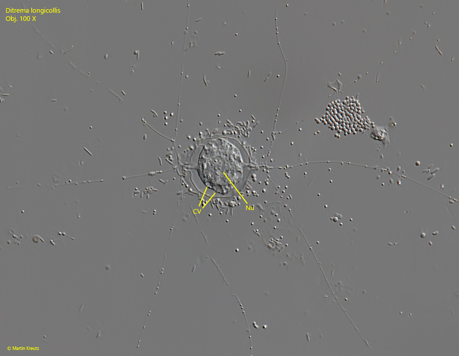
Fig. 1:Ditrema longicollis. L = 26 µm (from neck to neck). A fully extended specimen. CV = contractile vacuoles, Nu = nucleus. Obj. 100 X.
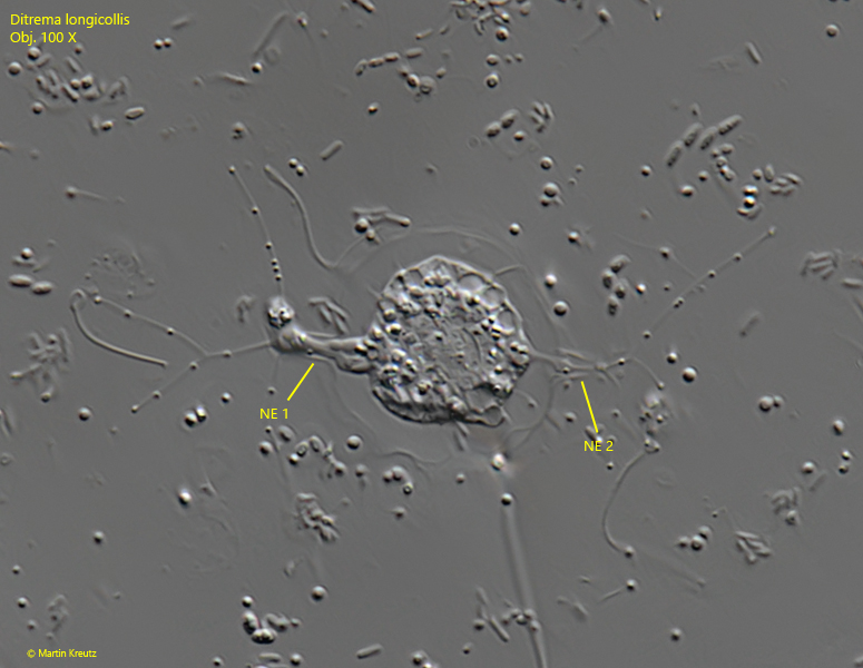
Fig. 2:Ditrema longicollis. L = 26 µm (from neck to neck). A second specimen with shorter granulofilopods. Note the two, opposite necks (NE 1, Ne 2). Obj. 100 X.
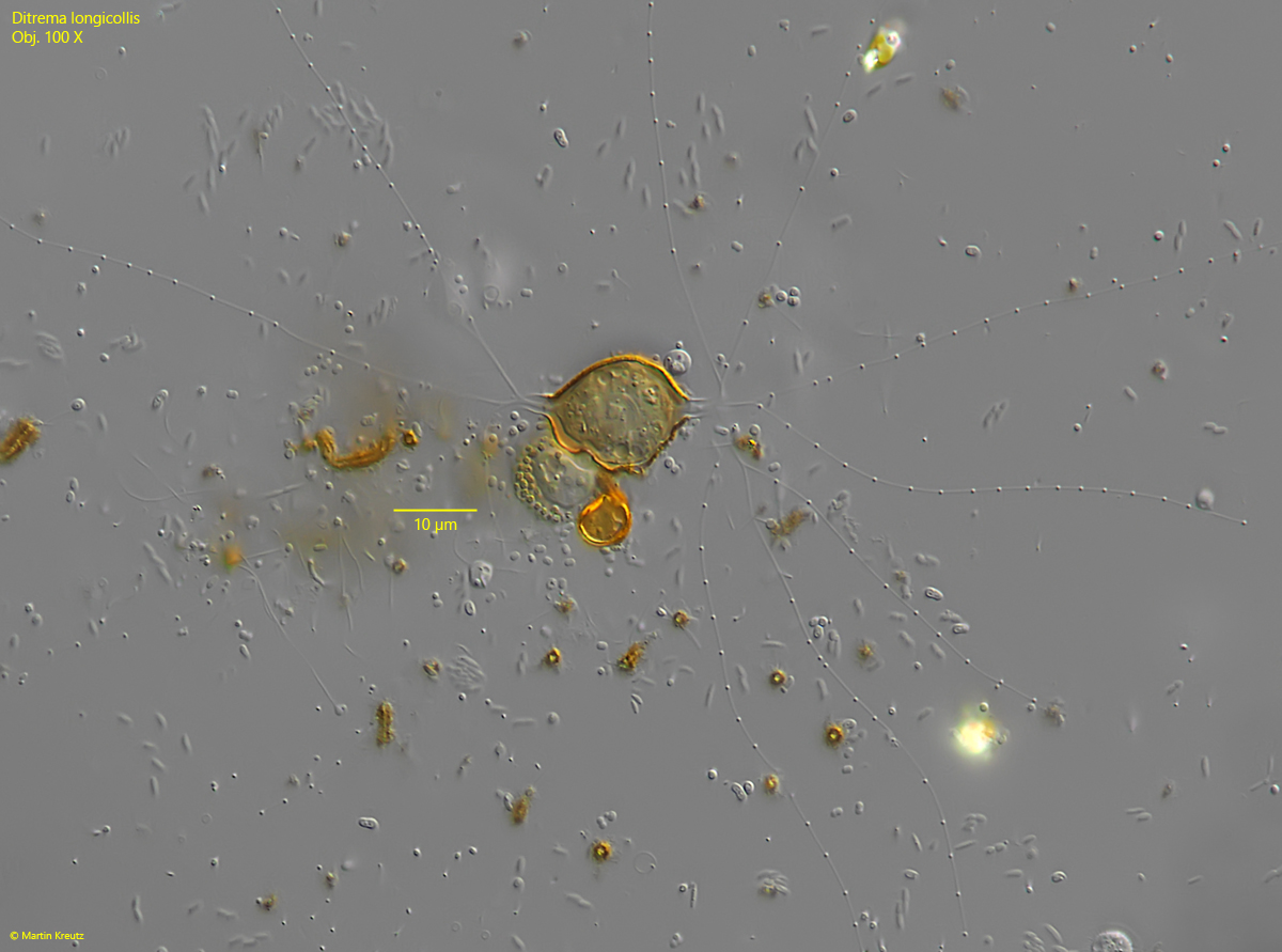
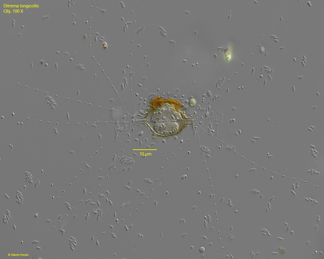
Fig. 3:Ditrema longicollis. L = 24 µm (from neck to neck). A third, fully extended specimen with an oval, slightly irregular shell. The shell of this older specimen is colored brown. Obj. 100 X.
Fig. 4:Ditrema longicollis. L = 24 µm (from neck to neck). A shell with two specimens after cell division. Obj. 100 X.