pharynx with a bundle of rod-shaped extrusomes, 13–15 µm long
also individual extrusomes in cytoplasm
12 rows of cilia per body side
contractile vacuole terminal
macronucleus moniliform, 5–10 nodules
ciliation soft and widely spaced
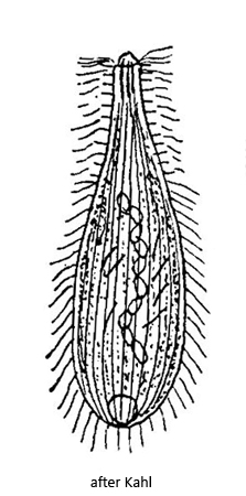
Enchelyodon monilatus
So far I have only been able to record Enchelyodon monilatus in the Simmelried, where I find it very rarely. Over a period of 20 years I have found only 5 specimens. I usually find the specimens in floating detritus flakes in old samples.
The elongated specimens in my population were 77–120 µm long, which is at the lower limit of the range of 120–170 µm given by Kahl. However, it also shows the high variability of the body length. The macronucleus is moniliform and consists in my specimens of 6–9 nodules. The micronuclei are not mentioned in Kahl’s decription, but in one of my examined specimens I could detect 2–3 of them (s. figs. 3 b and 5). Kahl describes the extrusomes as rod-shaped with a length of 13–15 µm. In my specimens they were slightly curved and 14–21 µm long. One end is clearly tapered, giving them a tusk-like appearance (s. figs. 3 c and 5). Kahl describes 12 rows of cilia on one side of the body with 12, which agrees exactly with my observations.
As there are no further findings and descriptions of Enchelyodon monilatus after Kahl, it is difficult to assess whether the characteristics of the specimens in my population are covered by Kahl’s brief description. A similar species Enchelyodon lageniformis was described by Vuxanovici in 1963, which is 80–120 µm long and has also a moniliform macronucleus consisting of 6 nodules. However, this species is not sufficiently characterized and possibly synonymous with Enchelyodon monilatus. Therefore I keep the classification Enchelyodon monilatus for the time being.
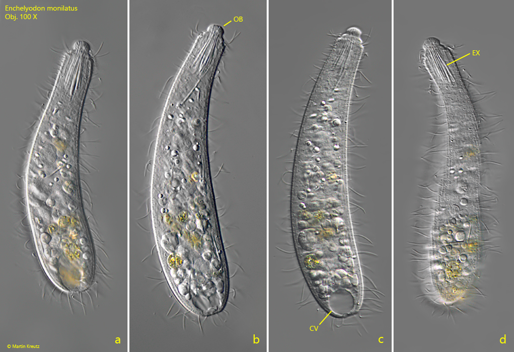
Fig. 1 a-d:Enchelyodon monilatus. L = 120 µm (of elongated specimen). Different focal planes of a freely swimming specimen. Note the narrow, hemispherical oral bulge (OB) and the bundle of extrusomes (EX) in the pharynx. CV = contractile vacuole. Obj. 100 X.
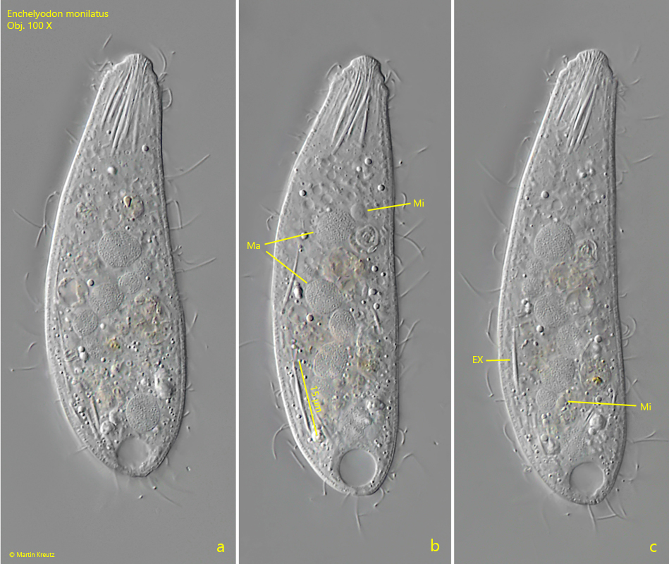
Fig. 2 a-c:Enchelyodon monilatus. L = 92 µm (of elongated specimen). A second freely swimming specimen. Note the nodules of the moniliform macronucleus (Ma) with the adjacent micronuclei (Mi). EX = extrusomes. Obj. 100 X.
Fig. 3 a-c:Enchelyodon monilatus. L = 101 µm. A third, fully elongated specimen. Note the two visible micronuclei (Mi) adjacent to the moniliform macronucleus. The slightly curved extrusomes (EX) of this specimen are 21 µm long. Obj. 100 X.
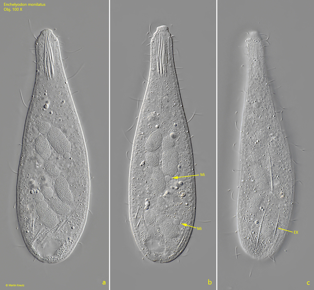
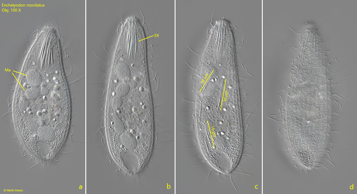
Fig. 4 a-d:Enchelyodon monilatus. L = 77 µm (of elongated specimen). A fourth, freely swimming specimen with 6 macronuclear nodules (Ma). The slightly curved and tusk-shaped extrusomes (EX) are 14–16 µm long. Obj. 100 X.
Fig. 5:Enchelyodon monilatus. A squashed specimen with three spherical micronuclei (arrows). Obj. 100 X.