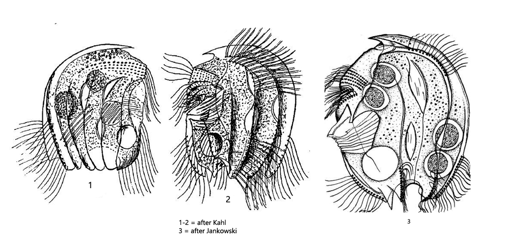
contractile vacuole below adoral zone of membranelles
Epalxella antiquorum
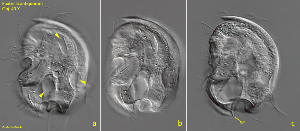
I have found Epalxella antiquorum in Simmelried only twice so far. First in March 2002 and then again in February 2017, so the species is quite rare. Therefore I have only comparatively few photos of it. The distinct identifying feature of Epalxella antiquorum is window-like openings in the pellicle on both sides of the body. They appear sharply outlined. According to Kahl, these are thinner areas of the pellicle. The species is described to have only blunt, rounded spines at the posterior end. However, in the specimen found in 2002 (s. fig. 1 a-c), I was able to document a rather sharp spine, localized slightly below the contractile vacuole (s. fig. 1c). This is not mentioned by any previous author. However, since it is the same specimen as shown in the figs. 1a and 1b (series of photos), a confusion with another species is excluded. I consider this a natural variety, as I could observe it also in other odontostomatid ciliates. The spine length can vary considerably within one species.
Fig. 1 a-c: Epalxella antiquorum. L = 73 µm. Three focal planes of the left side. Note the “windows” (arrowsheads), what are thin areas of the pellicle and the posterior spine (SP) . Obj. 40 X.
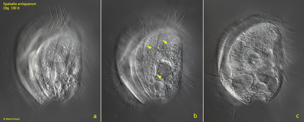
Fig. 2 a-c: Epalxella antiquorum. L = 59 µm. Three focal planes of the right side. Note the “windows” (arrowsheads), what are thin areas of the pellicle. Obj. 100 X.
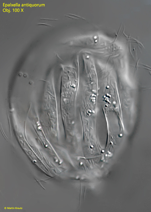
Fig. 3: Epalxella antiquorum. L = 76 µm. A further specimen from the right side. The pellicle is strongly grooved. Obj. 100 X.