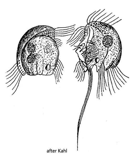
C-shaped ridge running in parallel to dorsal edge on right side
one long cirrus at posterior end of left side
elongated dorsal cilia
frontal tooth inconspicuous
single macronucleus and micronucleus
Epalxella exigua
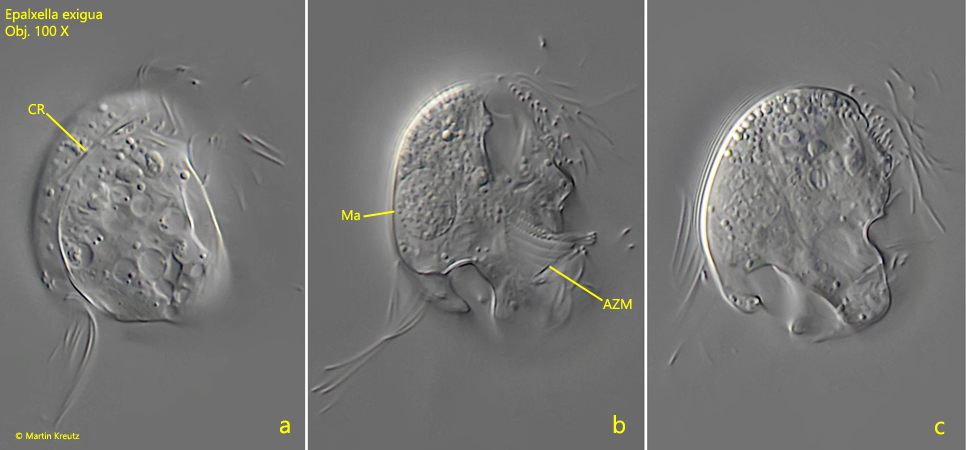
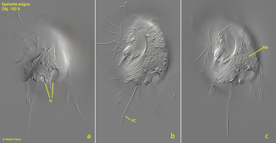
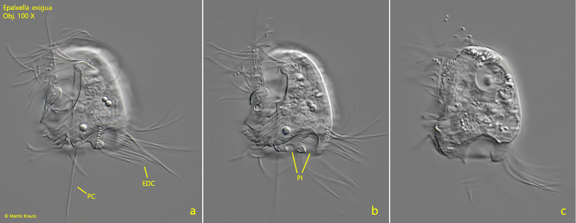
I find Epalxella exigua rarely and sporadically in the mud of Simmelried. I found specimens in January 2009 (s. fig. 3a-c) and in May 2021 (s. fig. 1a-c and fig. 2a-c). I recognize Epalxella exigua mainly by the C-shaped furrow on the right side (s. fig. 1a), the long posterior cirrus on the left side at the hind end (s. fig. 2b and fig. 3a) and the two indentations at the posterior end of the left side (s. fig. 2a and fig. 3b).
Fig. 1 a-c: Epalxella exigua. L = 28 µm. Lateral view of the right side of a slightly squashed specimen. Note the C-shaped ridge (CR) running in parallel to the dorsal edge on the right side. AZM = adoral zone of membranelles. Ma = macronucleus. Obj. 100 X.
Fig. 2 a-c: Epalxella exigua. L = 28 µm. Lateral view of the left side of a slightly squashed specimen. Note the two indentations at posterior end of the left side (PI). Ma = macronucleus, PC = posterior cirrus. Obj. 100 X.
Fig. 3 a-c: Epalxella exigua. L = 26 µm. Lateral view of a second specimen. EDC = elongated dorsal cilia, PI = posterior indentations, PC = posterior cirrus. Obj. 100 X.