extended zooids irregularly shaped, often roughly sigmoidal
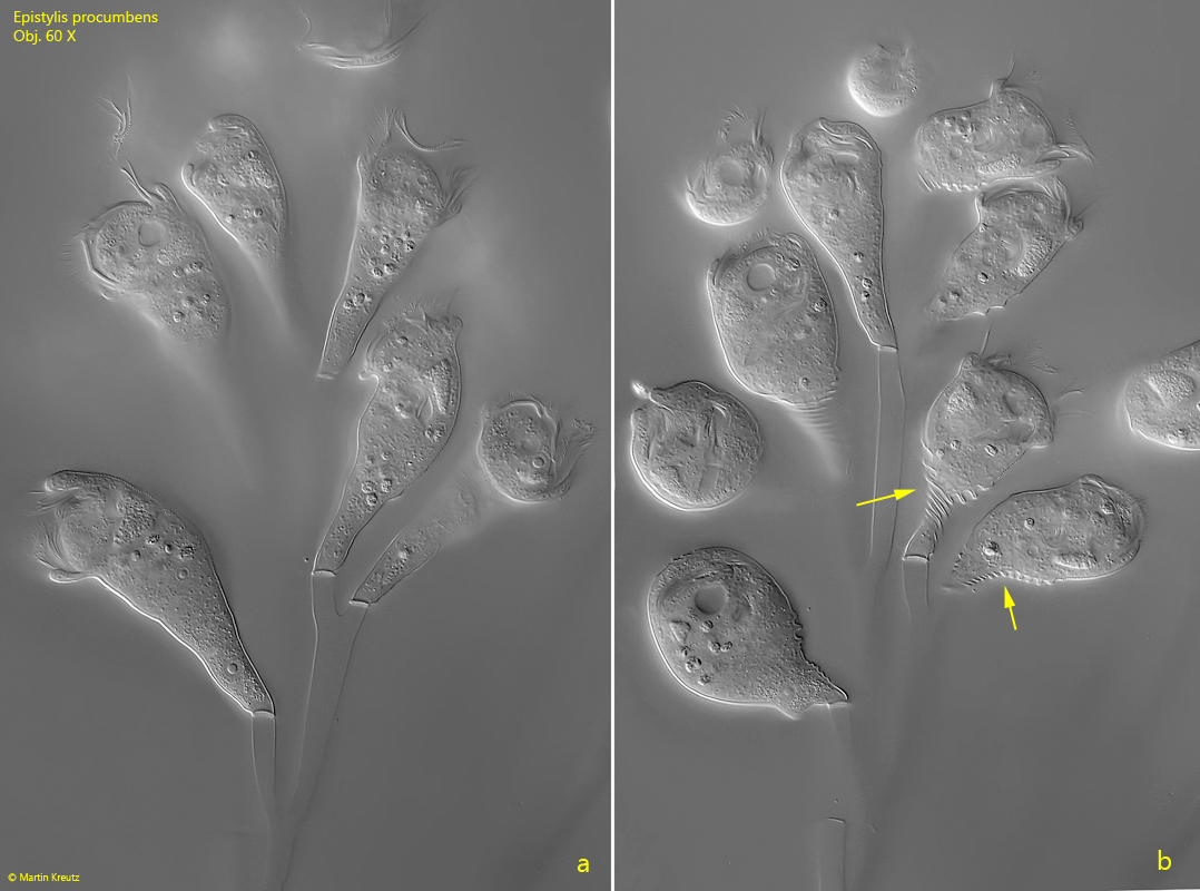
contracted zooids pyriform and distinctly annulated in posterior half
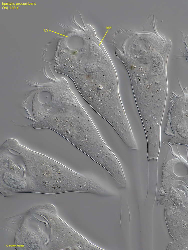
macronucleus reniform to semicircular in anterior half
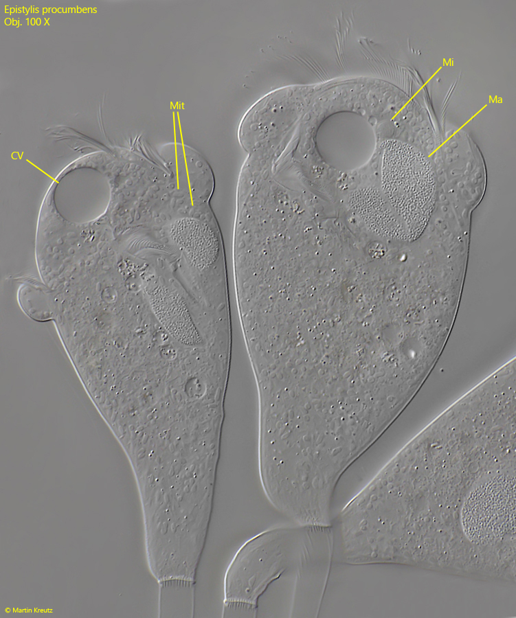
one micronucleus at anterior end of macronucleus
one contractile vacuole near peristomial collar
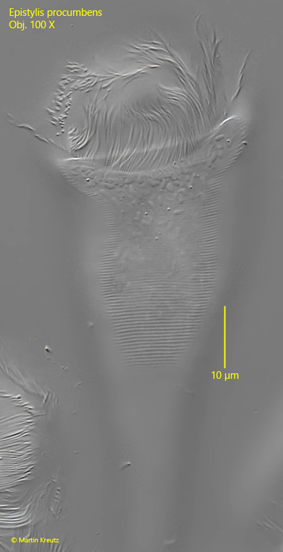
pellicle with about 137 transverse striae
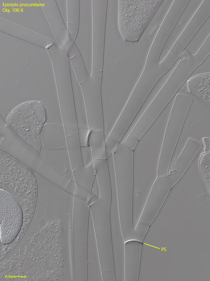
stalk non-contractile, dichotomously branched
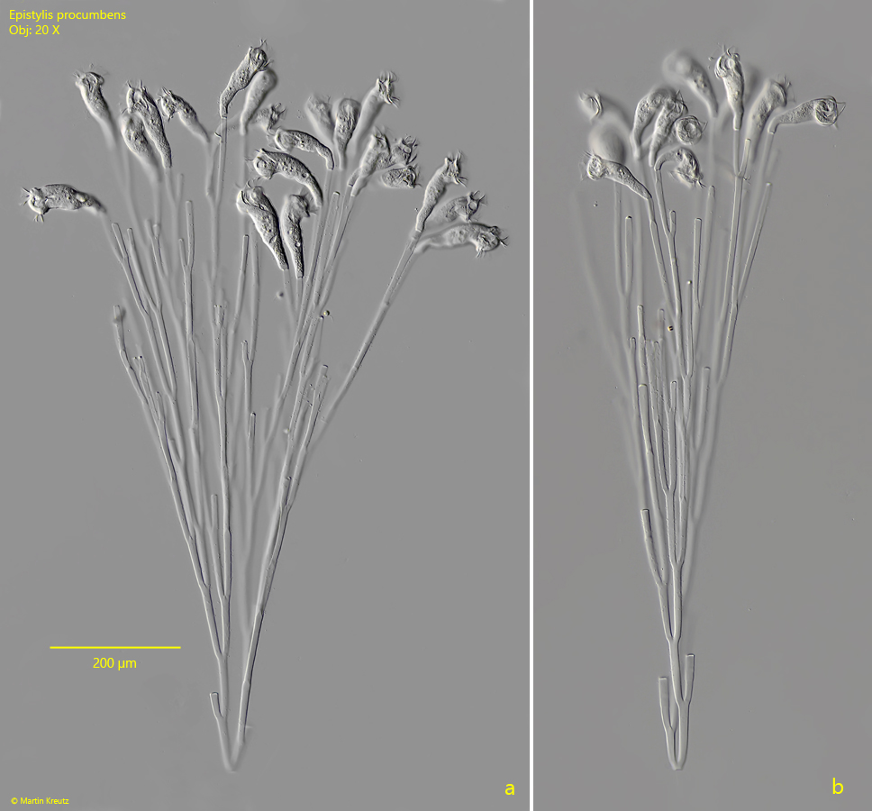
colonies up to 1 mm long
daughter colonies are separated at prospective separation sites
Epistylis procumbens
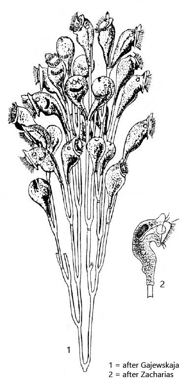
I regularly find the planktonic, peritrichous ciliate Epistylis procumbens in Lake Constance as well as in the Mühlhalden pond. The up to 1 mm long, slowly swimming colonies can be recognized even at very low magnification (s. fig. 1 a-b). A typical feature are the “tilted heads” of the colonies, because the zooids are bent sideways away from the longitudinal axis of the colony. In addition, the colonies are noncontractile and the stalks are dichotomously branched. Below the branching points, one often sees highly refractive separations (s. fig. 7). This is a kind of pre-determined breaking point at which daughter colonies “break off” and can be separated.
Fig. 1 a-b: Epistylis procumbens. Two freely swimming colonies. The colonies are 875 µm long (a) and 840 µm (b). Obj. 20 X.
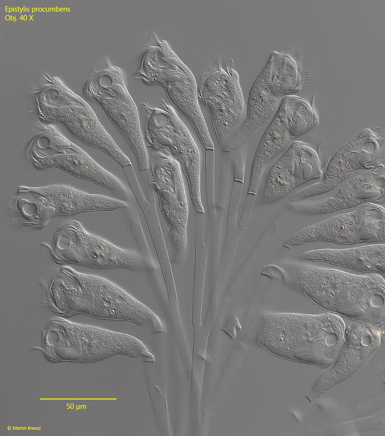
Fig. 2: Epistylis procumbens. L= 91–102 µm (of zooids). Some zooids of a slightly squashed colony. Obj. 40 X.
Fig. 3 a-b: Epistylis procumbens. L= 94–98 µm (zooids). A colony with extended (a) and contracted zooids (b). Note the annulated posterior portion of the contracted zooids (arrows). Obj. 60 X.
Fig. 4: Epistylis procumbens. L = 92–97 µm (zooids). In the slightly squashed specimens the single contractile valuole (CV) as well as the reniformed macronucleus (Ma) in the anterior half is visible. Obj. 100 X.
Fig. 5: Epistylis procumbens. In a more strongly squashed specimen the micronucleus (Mi) at the anterior end of the macronucleus (Ma) is visible. The 1–2 µm long “bubbles” are the mitochondria (Mit). CV = contractile vacuole. Obj. 100 X.
Fig. 6: Epistylis procumbens. The transverse striation of the pellicle. There are 17 lines per 10 µm. Obj. 100 X.
Fig. 7: Epistylis procumbens. Details of the stalks. Daughter colonies can be separated from the parent colony at the prospective separation sites (PS). Obj. 100 X.