1-3 pyrenoids per chloroplast, often covered with starch grains
spherical central nucleus
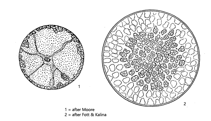
within cell radial strands of cytoplasm
chloroplasts arranged in a thin spherical layer of cytoplasm
Eremosphaera viridis
This large alga is mainly found in boggy waters, with a low pH. In Simmelried it was quite common until about 2010. After that the population been shrinking more and more, which is probably related to the increasing siltation of the area. Nevertheless, I regularly find Eremosphaera viridis in the samples, mostly among floating plants or in decomposing plant masses from the bottom of the water body. Because of the size of the cells (diameter mostly around 150 µm) and the radial plasma strands (s. fig. 1) it is easy to identify.
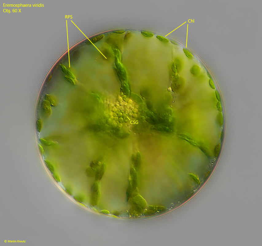
Fig. 1: Eremosphaeria viridis. D = 140 µm. Focus on the equatorial plane of a slightly squashed specimen. In the center of the cell an accumulation of starch grains is visible (CSG). The chloroplasts (Chl) can move in the radial plasm strands (RPS) from the periphery to the center of the cell. Obj. 60 X.
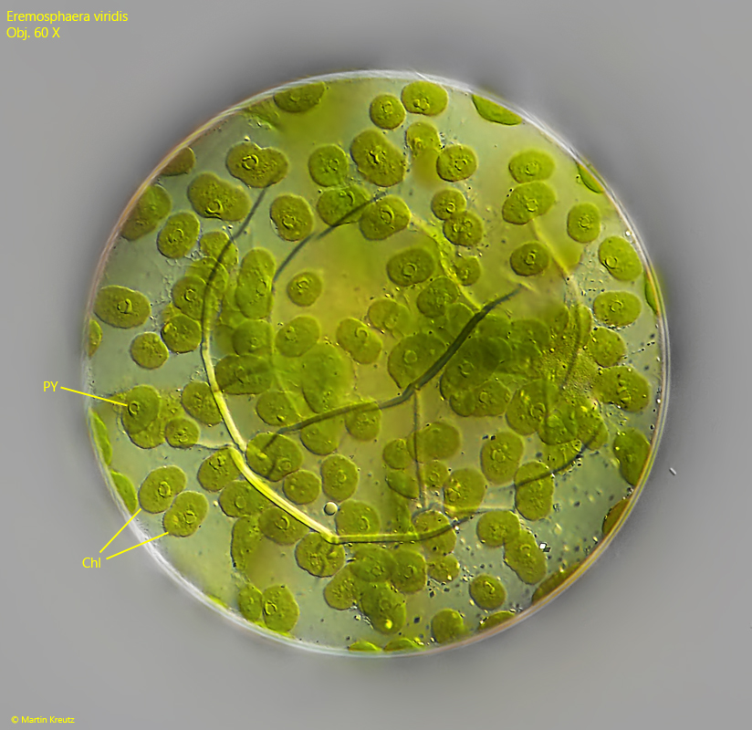
Fig. 2: Eremosphaeria viridis. D = 140 µm. Focus on the periphery of a slightly squashed specimen, where the chloroplasts (Chl) are arranged. In each chloroplast a small pyrenoid (PY) is visible covered by small starch grains. The “cracks” in the cell is the folded cell wall caused by the pressure of the coverslip. Obj. 60 X.
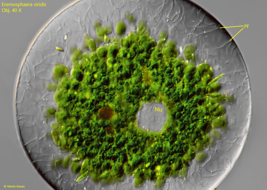
Fig. 3: Eremosphaeria viridis. D = 135 µm. A more strongly squashed specimen offers a view of the centrally located nucleus (Nu). The chloroplasts have all retracted to the center of the cell, caused by mechanical stress due to the thin water layer under the coverslip. Fine filaments of plasm (PF) can be seen in the outer layer. Obj. 40 X.
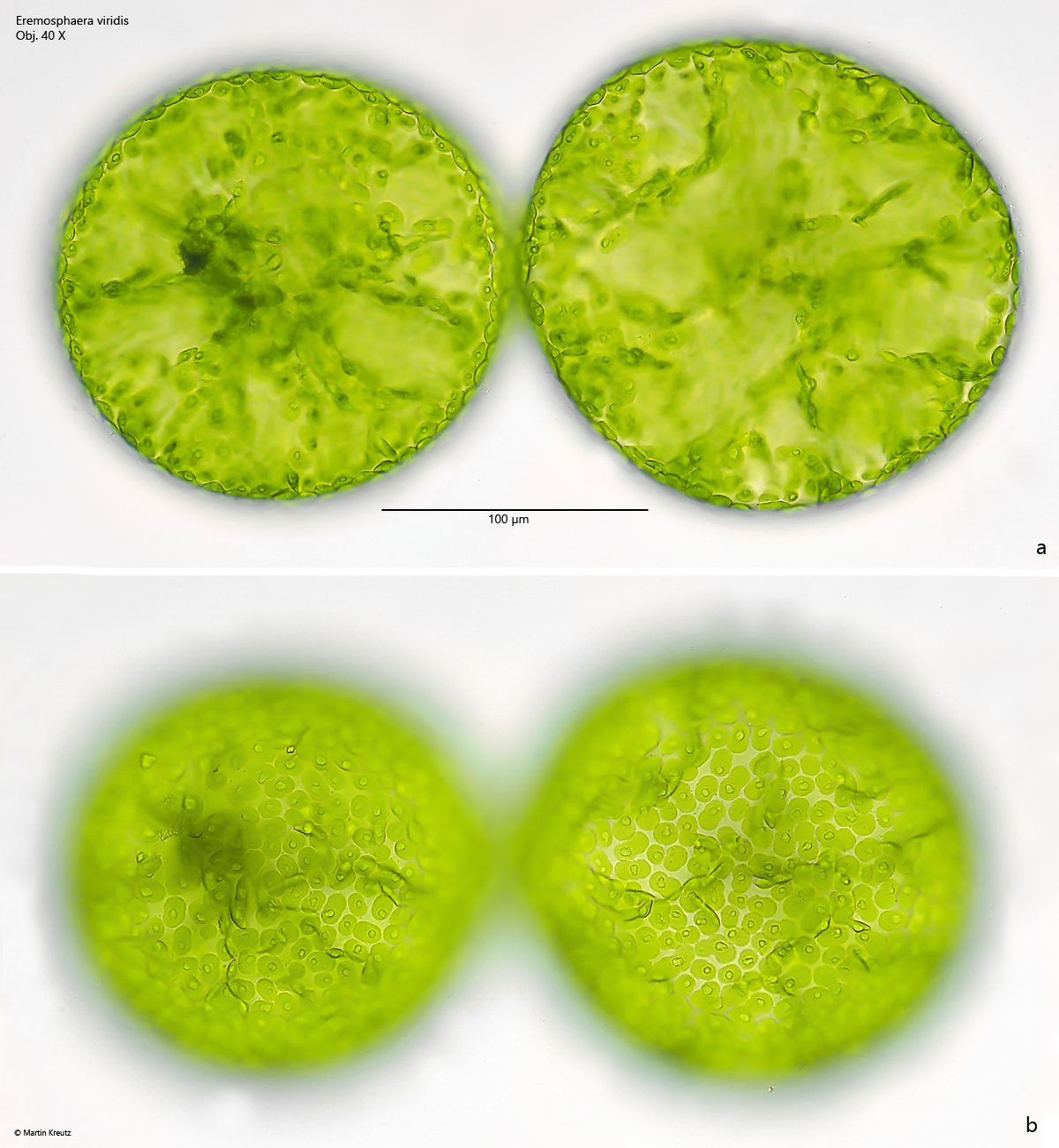
Fig. 4 a-b: Eremosphaeria viridis. D = 180 – 200 µm. Two focal planes of two slightly squashed cells in brightfield illuminaton. Obj. 40 X.
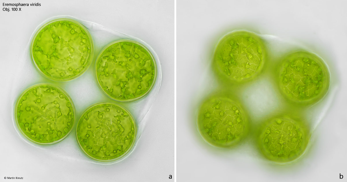
Fig. 5 a-b: Eremosphaeria viridis. An autospore with 4 daughter cells. Obj. 100 X.
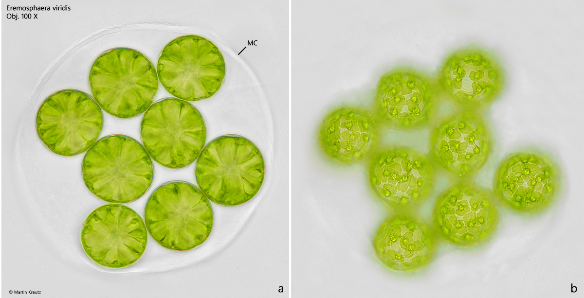
Fig. 6 a-b: Eremosphaeria viridis. A second autospore with 8 daughter cells enclosed in the cell wall of the mothercell (MC). Obj. 100 X.