stomach brownish or greenish from ingested cryptomonads
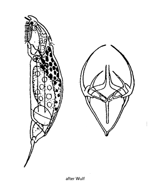
Erignatha clastopis
So far I have found Erignatha clastopis exclusively in the Simmelried. Mostly in floating plants or between decomposing plant masses. I recognize Erignatha clastopis mainly by the large, usually brown or greenish colored stomach (s. fig. 2 a-b), the thin, ventrally curved toes (s. fig. 1a) and the conspicuous crystalline inclusions in the gastric glands (s. figs. 3 a-b and 4). These crystalline inclusions light up very brightly in DIC, and are therefore quickly identifiable. The nature and function of these inclusions is unknown to my knowledge, except that they are a crystalline mass. Erighata clastopis feeds primarily on cryptomonads, which is why the stomach also shines brightly in DIC, due to undigested Maupas’ bodies of the cryptomonads.
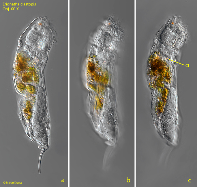
Fig. 1 a-c: Erignatha clastopis. L = 195 µm (inclusive toes). Lateral view of a freely swimming specimen. Note the crystalline inclusions (CI) in the gastric glands. Obj. 60 X.
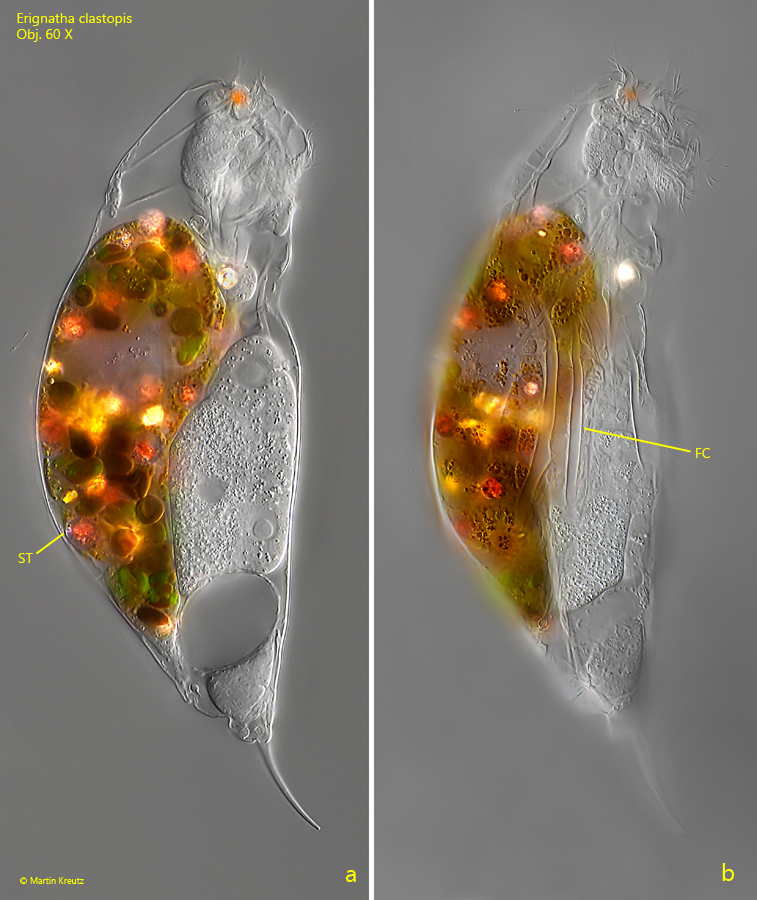
Fig. 2 a-b: Erignatha clastopis. L = 223 µm (including toes). Lateral view from right of a slightly squashed specimen. The crystalline inclusions in the gastric glands are highly birefringent and light up very brightly in DIC. The stomach (ST) of this specimen is completely filled with the remains of ingested cryptomonads. The Maupas’ bodies of the cryptomonads cause the shining fragments. FC = folded cuticula. Obj. 60 X.
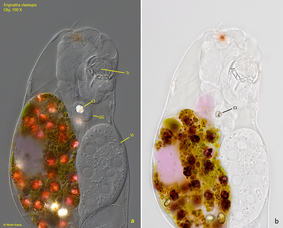
Fig. 3 a-b: Erignatha clastopis. A strongly squashed specimen in DIC (a) and brightfield illumination (b). Despite an underexposure of the DIC image the crystalline inclusion (CI) of the grastric glands (GG) lights up strongly and no details can be recognized. In brightfield illumination the inclusion appears brownish and seems to consist of radially grown crystals emanating from a central nucleus of crystallization. Tr = trophi, Vi = vitellarium. Obj. 100 X
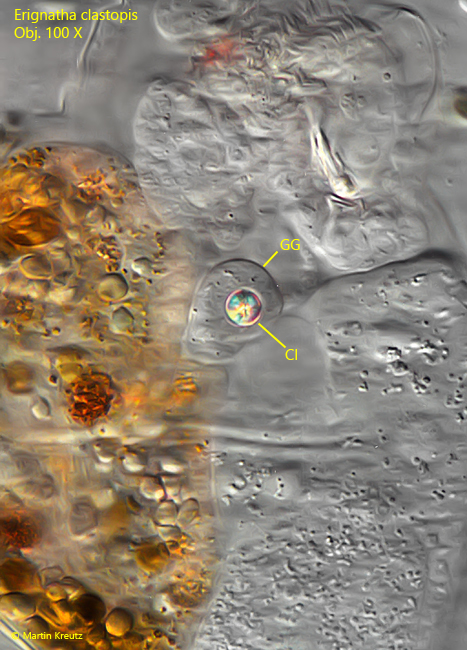
Fig. 4: Erignatha clastopis. This image was taken with a strong underexposure for visualisation of the details in the crystalline inclusion (CI) of the gastric glands (GG) in DIC. The inclusion appears as a spherule colored by the interference colors due to its birefringent properties. Obj. 100 X
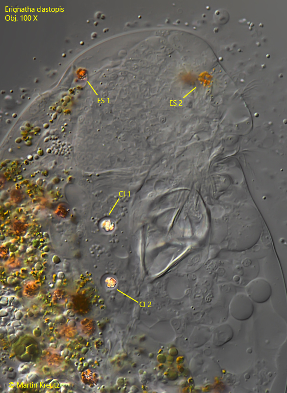
Fig. 5: Erignatha clastopis. In this strongly squashed specimen the pair of eyespots is visible (ES 1, ES 2) and the pair of crystalline inclusions (CI 1, CI 2) of the two gastric glands. Obj. 100 X