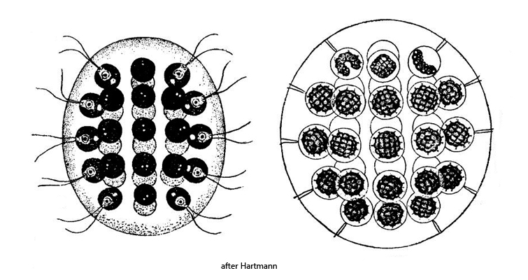
colony ellipsoidal or spherical with a mucilaginous envelope
length 50–200 µm
colony consisting of 4–8–16–32–64 cells (in most cases 32)
cells arranged in 5 layers (4–8–8–8–4)
cells spherical, sub-spherical or pear-shaped each with 2 flagella of equal length
flagella cross the mucilaginous envelope through canals
each cell with 2 contractile vacuoles
one chloroplast, cup-shaped
one pyrenoid
one eyespot
Eudorina elegans
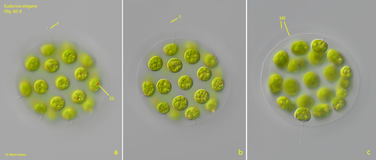
Eudorina elegans is a widespread volvococcal alga, which sometimes occurs in masses especially in my sampling site Simmelried. The spherical colonies with mostly 32 cells are easy to identify (s. figs. 1 a-c and 2 a-b).
Eudorina elegans can reproduce asexually by vegetative division, but also by sexual reproduction, as there are male as well as female colonies. In the vegetative state the sexes cannot be distinguished. Only at stages of reproduction it is possible to determine which sex is present. In the male colonies clusters of sperm cells forme (s. figs. 6 and 7) and in the female colonies either immobile aplanospores or flagellated zoospores are formed after fertilization. When immobile aplanospores are formed, they begin to germinate and form new colonies by cell division (s. fig. 8).
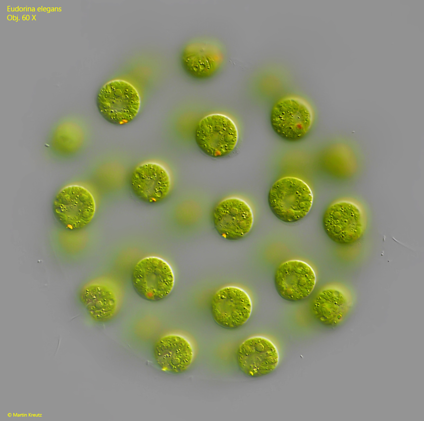
Fig. 1 a-c:Eudorina elegans. D = 132 µm (of colony). The focal planes of a freely swimming colony. Note the eyespots (ES) of the cells. F = flagella, ME = mucilaginous envelope. Obj. 60 X.
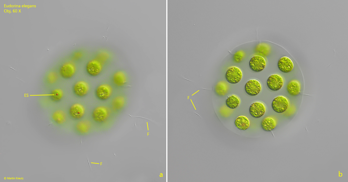
Fig. 2 a-b:Eudorina elegans. D = 98 µm (of colony). A second freely swimming colony. F = flagella, ES = eyespots. Obj. 60 X.
Fig. 3:Eudorina elegans. D = 131 µm (of colony). The cells of a colony in detail. Note the contractile vacuoles (CV) and the eyespot (ES) of some spezialized cells. Obj. 100 X.
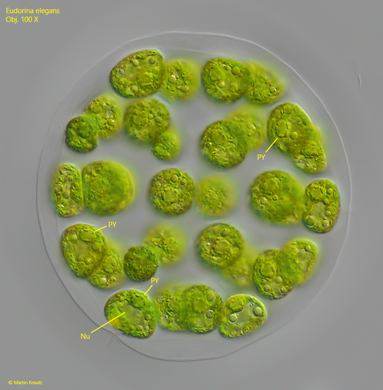
Fig. 4:Eudorina elegans. D = 131 µm (of colony). Focal plane on cells with a visible pyrenoid (PY) and the centrally located nucleus (Nu). Obj. 100 X.
Fig. 5:Eudorina elegans. D = 153 µm (of colony). Focal plane on cells with an eyespot. Obj. 60 X.
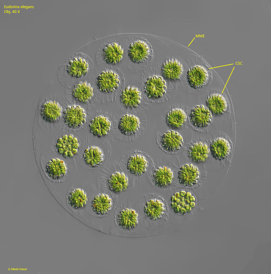
Fig. 6:Eudorina elegans. The formation of clusters of sperm cells (CSC) in a male colony. MME = mucilaginous envelope of mother cell. Obj. 40 X.
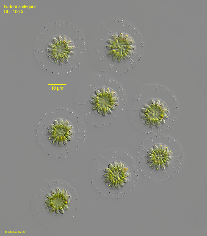
Fig. 7:Eudorina elegans. The released clusters of sperm cells. Obj. 100 X.
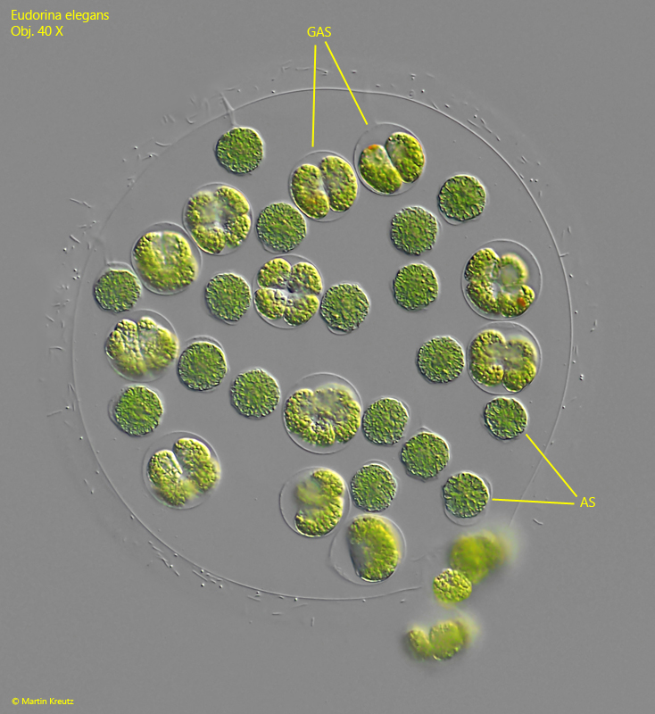
Fig. 8:Eudorina elegans. Aplanospores (AS) and germinating aplanospores (GAS) in a female colony. Obj. 40 X.