cell cylindrical with rounded anterior and posterior end
length 35–50 µm, width about 13 µm
two ribbon-shaped chromatophores with length nearly of the cell
no pyrenoids
paramylon bodies roundish, oblong or short cylindrically
eyespot roundish or slightly angular
flagellum shorter than body length
nucleus located between the chromatophores in posterior half of cell
metabolic movement with the rounded form of the posterior end retained troughout
Euglena van-goori
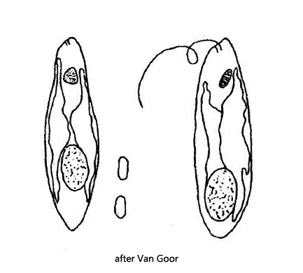
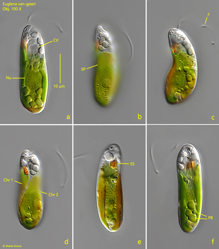
Euglena van-goori was first described in 1925 by Van Goor as Euglena obtusa. Since this name was already used in 1884 by Schmitz, Deflandre renamed it in 1928 to Euglena van-goori. Obviously only the original description of this Euglena species by Van Goor is available. Further finds or descriptions do not seem to exist. For the identification the number and shape of the chromatophores is important. I could recognize two ribbon-shaped chromatophores, which almost reached the length of the entire cell (s. figs. 1d and 1e). Pyrenoids are not present. Thus it could only be Euglena van-goori, because the similar species Euglena bivittata has also two chromatophores, but these reach only half of the cell length and are located above and below the nucleus. There is no other alternative Euglena species with two ribbon-shaped chromatophores.
My specimen was only 28 µm long and thus smaller than indicated by Van Goor (35–50 µm). However, since there are no other observations and measurements of this species besides the description by Van Goor, this length may still be within the range of variantion. All other characteristics agree with the description of Van Goor. I could also see a delicate striation of the pellicle (s. figs. 1b and 2b) running counterclockwise across the body, which Van Goor did not mention. I found the specimen shown in figs. 1a-f and fig. 2a-b in Simmelried in February 2023. I may have overlooked Euglena van-goori in previous samples because of its small size.
Fig. 1 a-f:Euglena van-goori. L = 28 µm. Different focal planes of a freely moving specimen. Chr 1, Chr 2 = chromatophores, CV = contractile vacuole, ES = eyespot, Nu = nucleus, PB = paramylon bodies, SP = striation of the pellicle. Obj. 100 X.
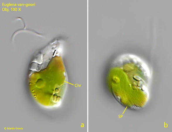
Fig. 2 a-b:Euglena van-goori. L = 28 µm. Focal plane on one of the chromatophores (a, Chr) and the striation of the pellicle (b, SP) of a slightly squashed and contracted specimen. Obj. 100 X.