oral aperture circular and surrounded by 8–11 apertural scales
pointed ends of apertural scales are thickened
nucleus globular in posterior third of cell
Euglypha filifera
I found Euglypha filifera in September 1997 and in August 2006 in the Simmelried. After that I have not registered any more finds.
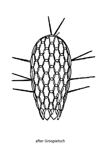
In Euglypha filifera the spines are arranged on the margin of the flattened shell (s. fig. 1 c), but only on the posterior two-thirds. The anterior third of the shell is free from spines as well as the lateral surfaces. The shell itself is composed of oval scales, which appear hexagonal because they overlap each other (s. fig. 2 b). The round mouth opening is surrounded by 8–11 specially shaped scales (s. fig. 3 a – c). At the pointed ends these apertural scales are thickened.
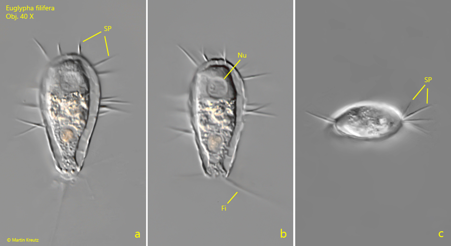
Fig. 1 a-c: Euglypha filifera. L = 53 µm (without spines). A freely moving specimen from lateral (a, b) and from apical (c). Note the lateral flattening of the shell and that the spines arise only on the margin (c). Fi = filopodia, Nu = nucleus, SP = spines. Obj. 40 X.
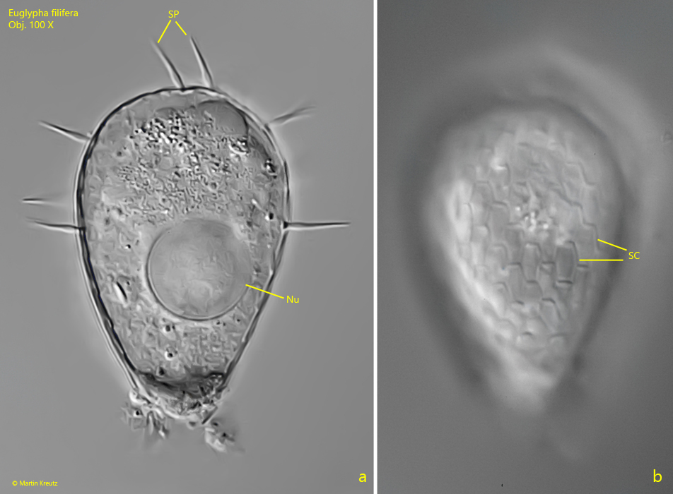
Fig. 2 a-b: Euglypha filifera. L = 54 µm (without spines). Two focal planes of a squashed specimen. Note the oval scales (SC) of the shell appearing hexagonal due to overlapping. Nu = nucleus. Obj. 100 X.
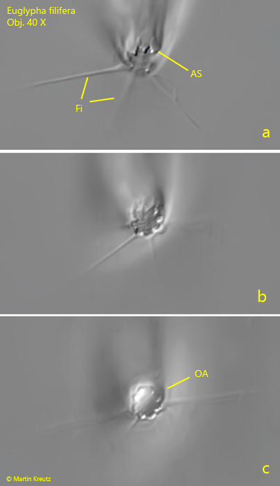
Fig. 3 a-c: Euglypha filifera. Apical view of a freely moving specimen. Note the apertural scales (AS) surrounding the circular oral aperture (OA). Obj. 40 X.