body oval, moderately flattened, in cross section elliptical
cytoplasm colorless, without pigment spot at anterior end
length 80–130 µm, width 60–100 µm
one contractile vacuole in mid-body on right side, with 2–4 excretion pores
macronucleus ellipsoid, about 24 x 15 µm with a globose micronucleus located in an indentation of macronucleus
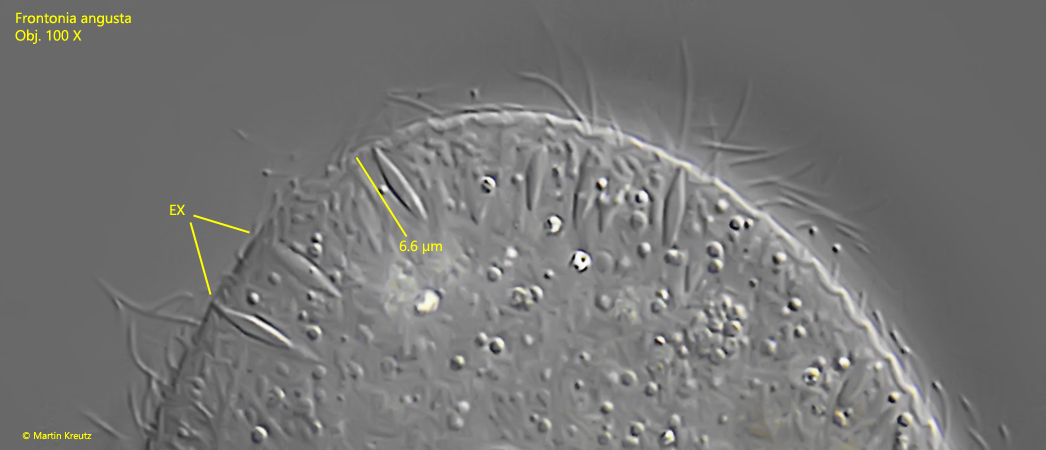
fringe of spindle-shaped extrusomes, about 6 µm long
80–105 longitudinal rows of cilia
tuft of elongated cilia at posterior end
pellicle with quadrangular pattern
Frontonia angusta
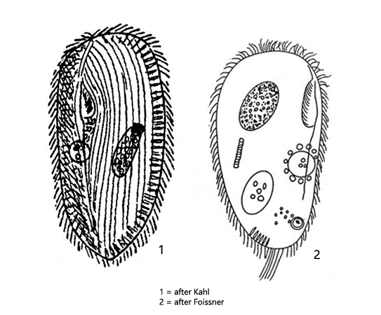
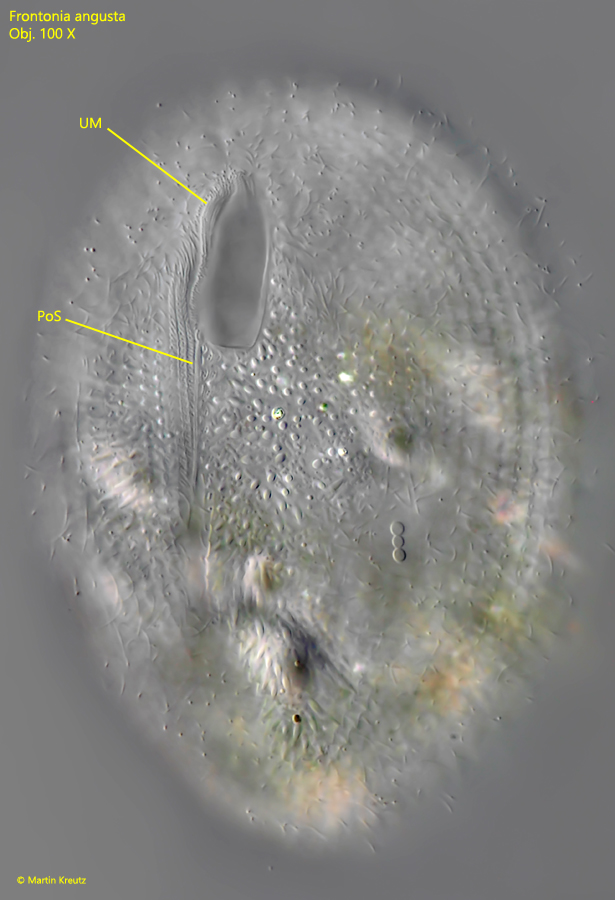
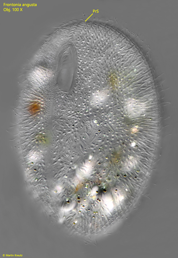
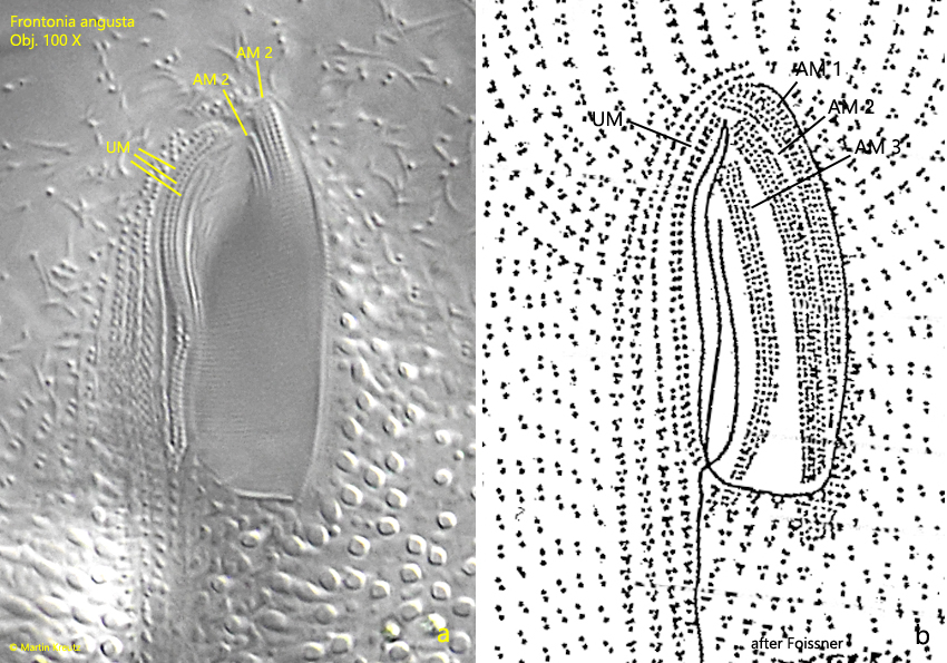
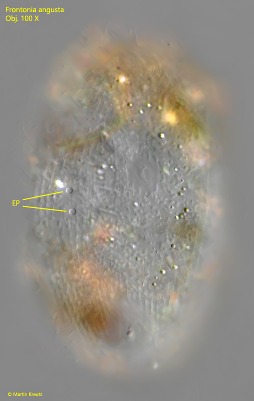
Frontonia angusta is very similar to Frontonia acuminata. However, both species can be distinguished by a dark pigment spot in the apical area, present only in Frontonia acuminata. Since I could not discover a pigment spot, Frontonia angusta (s. fig. 1-b) must be present here. In Frontonia angusta a rather large micronucleus is attached to the macronucleus and located in an indentation of the macronucleus (s. fig. 2). There is only one contractile vacuole, which is located in the middle of the body on the right side (s. fig. 2). It should have 2–4 excretory pores. In my specimens there were always two (s. fig. 7). Since the number is variable in this species, it cannot be used as an identifier, but rather as an indication. However, there are also genera where the number of excretory pores is constant (e.g. Paramecium) and can be used as an identification characteristic. With higher magnification one recognizes the typical features of the mouth organization of Frontonia. The undulating membranelles and two of the three adoral membranelles can be seen (s. fig. 6 a-b). However, there is a special feature in Frontonia angusta, which interestingly is not mentioned by Foissner, but Kahl described it: “…hinter dem Mund, links von der Naht ein kleines dreiseitiges Feld größerer, stark gerahmter Wimpernfeldchen (behind the mouth, to the left of the postoral suture a small three-sided field of framed cilia is visible)”. In fact, this distinctly shaped field is very conspicuous (s. fig. 3 a-b). The purpose of this trigonal field remains uncertain. In Frontonia, there is a so-called preoral suture and a postoral suture. Both can only be seen when carefully focusing on the ventral side (s. figs. 4 and 5). The postoral suture is shaped like a groin and is supposed to be a kind of predetermined breaking point of the pellicle according to Kahl. If very large food is ingested, the mouth opening along the suture can widen like a gap but I have never observed this process myself.
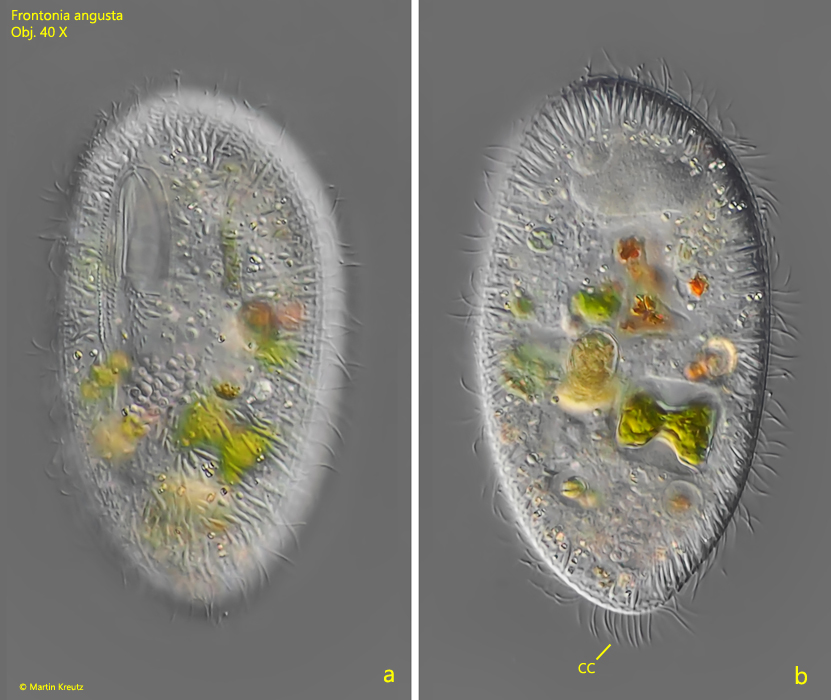
Fig. 1 a-b:Frontonia angusta. L = 110 µm. Two focal planes of a freely swimming specimen. CC = tuft of caudal cilia. Obj. 40 X.
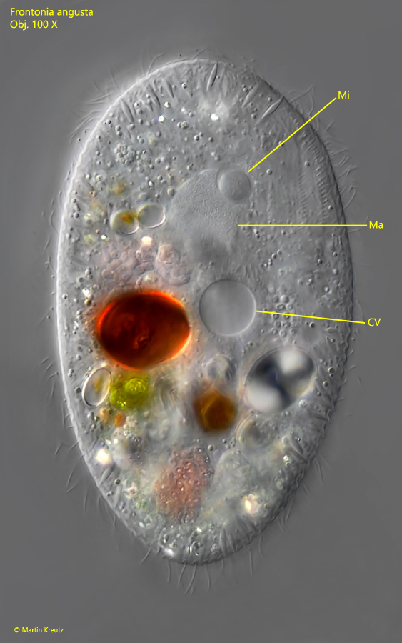
Fig. 2:Frontonia angusta. L = 105 µm. A slightly squashed specimen from right. CV = contractile vacuole, Ma = macronucleus, Mi = micronucleus. Obj. 100 X.
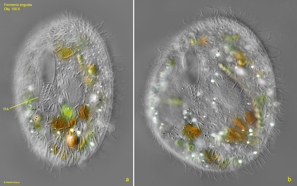
Fig. 3 a-b:Frontonia angusta. Focal plane on the triangular field (TFA) below the mouth opening during reduction of the layer thickness. Obj. 100 X.
Fig. 4:Frontonia angusta. L = 118 µm. Focal plane on postoral suture (PoS). UM = undulating membranelles. Obj. 100 X.
Fig. 5:Frontonia angusta. L = 118 µm. The same specimen shown in fig. 3 with focal plane on the praeoral suture (PrS). Obj. 100 X.
Fig. 6 a-b:Frontonia angusta. Details of the mouth opening (a) compared to a drawing after Foissner (b). UM = undulating membranelles, AM 1–3 = adoral membranelles. Obj. 100 X.
Fig. 7:Frontonia angusta. Two excretion pores (EP) of the contractile vacuole on the right side in the middle of the cell. Obj. 100 X.
Fig. 8:Frontonia angusta. The spindle-shaped extrusomes (EX) are about 6 µm long. Obj. 100 X.