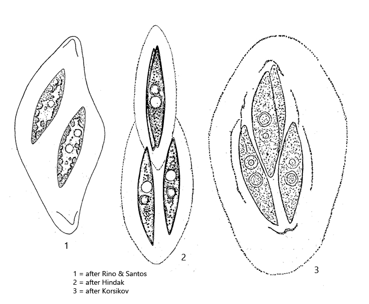
colonies of 2-4-8-cells in a mucilaginous envelope
cells spindle-shaped, 25 – 54 µm long
cells arranged serially and shifted against each other
many parietal chloroplasts, plate-shaped
nucleus central
one or two pyrenoids
cells often filled with starch grains
Fusola viridis
I find Fusola viridis exclusively in Simmelried in decomposing plant masses, but always sporadically. The last finds were in April 2016 and in September 2022. This alga can very easily be confused with Elakatothrix, which also has spindle-shaped cells in a mucilage shell. However, members of the genus Elakatothrix have only 1 or 2 chloroplasts.
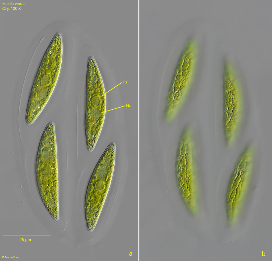
Fig. 1 a-b: Fusola viridis. L = 49 -55 µm. Two focal planes of a slightly squashed colony of 4 cells. Nu = nucleus, PY = pyrenoid. Obj. 100 X.
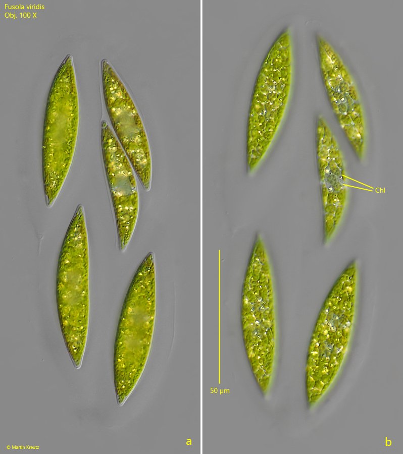
Fig. 2 a-b: Fusola viridis. L = 36 -59 µm. Two focal planes of a second colony with 5 cells. One cell of the colony had recently divided. The plate-shaped chloroplasts are visible in the two daughter cells (Chl, s. fig. 2b). Obj. 100 X.
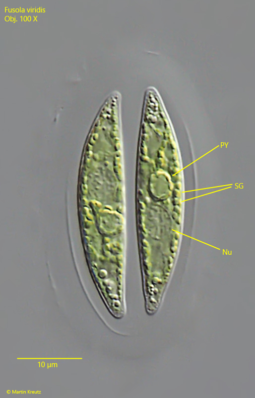
Fig. 3: Fusola viridis. L = 35 – 37 µm. A young colony of 2 cells. Nu = nucleus, PY = pyrenoid, SG = starch grains. Obj. 100 X.