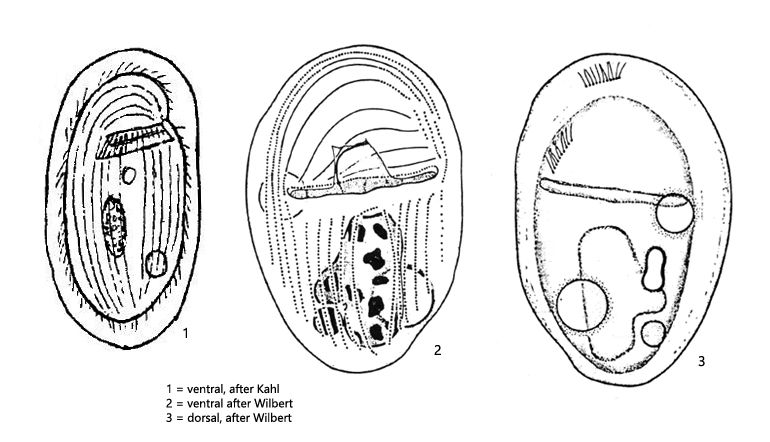
body ellipsoid, dorso-ventrally flattened, left margin slightly less convex than right
length 50–70 µm, width 26–32 µm
macronucleus ellipsoid, in its center a nucleolus, one spherical micronucleus directly adjacent
near macronucleus a light-refractive structure of unknown function
two contractile vacuoles in diagonal position
on the posterior half of the ventral side a non-ciliated central field
mouth opening a transverse slit, slightly anterior to the cell center
circumoral cilia forming a lamellar structure
dorsal brush consists of two rows of cilia, which are arranged almost perpendicular to each other
Gastronauta membranaceus
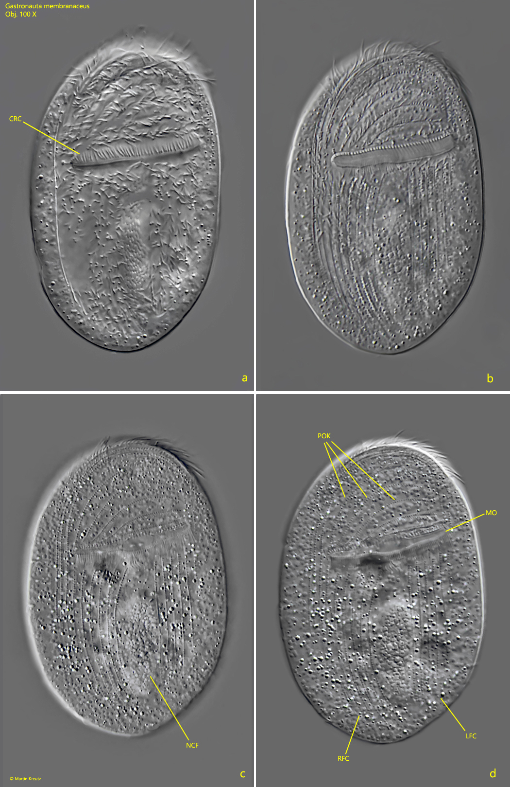
Gastronauta membranaceus belongs to the cyrtophorid ciliates. The genera Chilodonella or Trithigmostoma also belong to this group. The affiliation to this group results from the type of ciliation. On the dorsal side they are (almost) naked, while the ventral side has stripe-like rows of cilia (kineties) in front of and behind the mouth opening. However, while Chilodonella and Trithigmostoma have a mouth opening with a pharyngeal basket, that of Gastronauta membranaceus is shaped as a transverse slit (s. fig. 1a-d). This is a useful adaptation to grazing on bacteria growing on surfaces.
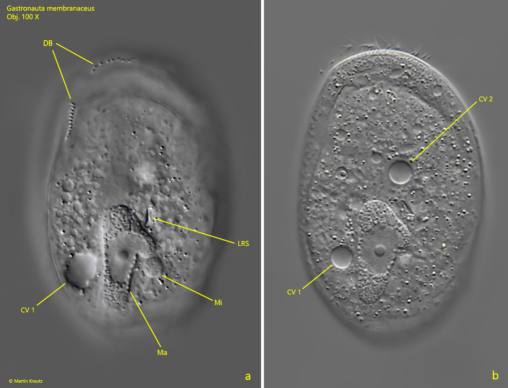
I have found Gastronauta membranaceus once so far (January 2019) and then only a few specimens. I found them on a floating coverslip, the underside of which they grazed. At low magnifications the specimens are very easy to confuse with Chilodonella. Perhaps this is because only a few finds have been reported. On the floating coverslip, the ventral side is always facing the observer. What is typical of Gastronauta membranceus is a non-ciliated field in the posterior, ventral half of the body (s. fig. 1c). In the similar species Gastronauta clatratus the entire posterior, ventral half of the body is ciliated. The dorsal side of Gastronauta mambranaceus is naked except for the dorsal brush, which consists of two short rows of cilia that are nearly perpendicular to each other (s. fig. 2a). Two contractile vacuoles are present, which are diagonal to each other, one anteriorly on the right and the other posteriorly on the left (s. fig. 2b). Near the macronucleus, an hourglass-shaped light-refractive structure is visible (s. fig. 2a). This is not phagocytosed food, but an organelle of unknown function.
Fig. 1 a-d:Gastronauta membranaceus. L = 64 µm. Different focal planes from ventral of a slightly squashed specimen. CRC = circumoral row of cilia, LFC = left field of cilia, MO = mouth opening, NFC = non-ciliated field, POK = pre-oral kineties, RFC = right field of cilia. Obj. 100 X.
Fig. 2 a-b:Gastronauta membranaceus. L = 64 µm. Dorsal view of a slightly squashed specimen seen from the ventral side. Note the light-refractive structure (LRS) of unknown function near the macronucleus. CV 1, CV 2 = contractile vacuoles, Ma = macronucleus, Mi = micronucleus, DB = dorsal brush. Obj. 100 X.