body is ovoid, spindle-shaped or “tailed” at the posterior end
oral aperture located in the anterior third is from “Glaucoma“-type with three distinct membranelles
CV central
macronucleus globular or oval, centrally located or in the anterior third
inconspicuous fringe of extrusomes present
Glaucoma frontata
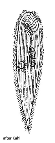
I found Glaucoma frontata for the first time in Simmelried in 1999. After that this species occured in intervals of several years. The identification of Glaucoma frontata according to the drawing and description of Kahl is difficult, because the shape of Glaucoma frontata can vary very much. There are varieties from slender spindle-shaped to broadly oval or with a distinctly tapered posterior end. Therefore, I provide some examples of freely swimming specimens with different shapes found between 2008 and 2019 (s. figs. 1 a-b to 6 a-d).
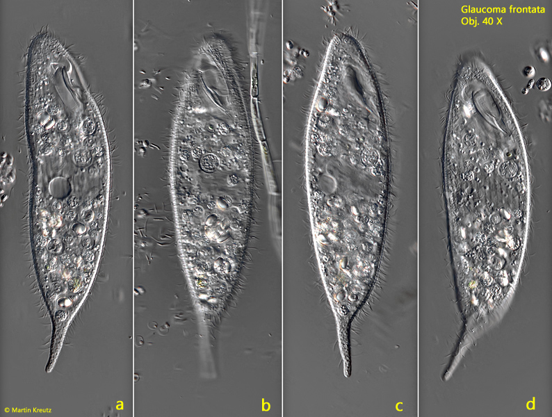
Fig. 1 a-d:Glaucoma frontata. L = 131 µm. Freely swimming specimen with a slightly tapered posterior end. Obj. 40 X.
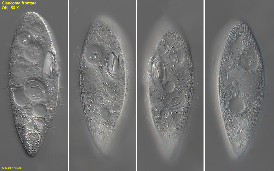
Fig. 2 a-c:Glaucoma frontata. L = 121 µm. A second freely swimming specimen with an almost ellipsoid shape. CV = contractile vacuole, Ma = macronucleus. Obj. 60 X.
Fig. 3 a-b:Glaucoma frontata. L = 121 µm. The slightly squashed specimen as shown in fig. 2 a-b. CV = contractile vacuole, Ma = macronucleus, ME = adoral membranelle, MO = mouth opening, UM = undulating membrane. Obj. 100 X.
Fig. 4 a-d:Glaucoma frontata. L = 133 µm. A freely swimming, spindle-shaped specimen. Obj. 60 X.
Fig. 5 a-d:Glaucoma frontata. L = 120 µm. Freely swimming specimen with a distinct “tail”. Obj. 40 X.
Fig. 6 a-d:Glaucoma frontata. L = 160 µm. A second freely swimming specimen with a distinct “tail”. Obj. 100 X.
Fig. 7 a-d:Glaucoma frontata. L = 160 µm. A third freely swimming specimen from dorsal with a distinct “tail”. Obj. 100 X.
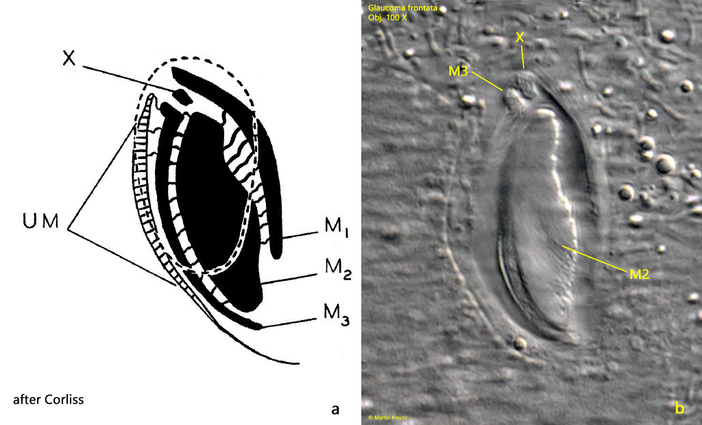
In a slightly pressed specimen, the structure of the oral aperture can be seen in more detail. The structure is of the typical Glaucoma style. Three adoral membranelles (M1-M3) are parallel and surrounded by an undulating membrane from the right side (s. fig. 8). The middle adoral membranelle (M2) is most pronounced, just as Corliss drew it (s. fig. 9 a).
Fig. 8:Glaucoma frontata. The oral apparatus with the three adoral membranelles (M1-M3). UM = undulating membrane. Obj. 100 X.
The ciliation of the oral aperture of Glaucoma frontata was described in detail by Corliss (s. fig. 9 a). According to his investigation, a small patch of cilia is located at the anterior end of the prominent adorale membranelle M2, which he called “enigmatic structure” or “X-body”. He suggested that it was a separate piece of the membranelle M2. This structure is difficult to see without silver impregnation. However, when the oral aperture is focussed from the dorsal side of the ciliate, it can be observed (s. fig. 9 b):
Fig. 9 a-b:Glaucoma frontata. a) the arrangement of the adoral membranelles (M1 – M3) and undulating membrane (UM) of the oral aperture according to Corliss including the enigmatic X-Body. b) focus on the oral aperture of Glaucoma frontata from dorsal. M1 – M 3 = adoral membranelles, UM = undulating membrane, X = X-body. Obj. 100 X.
Fig. 10:Glaucoma frontata. The mouth opening of a second specimen in detail. M2 + M3 = adoral membranelles, UM = undulating membranene. Obj. 100 X.
Fig. 11:Glaucoma frontata. The macronucleus (Ma) and micronucleus (Mi) in a squashed specimen. FV = food vacuoles, ME = adoral membranelle, UM = undulating membranene. Obj. 100 X.
Fig. 12:Glaucoma frontata. The the rod-shaped extrusomes (arrows) with a length of 1.8–2.0 µm. Obj. 100 X.