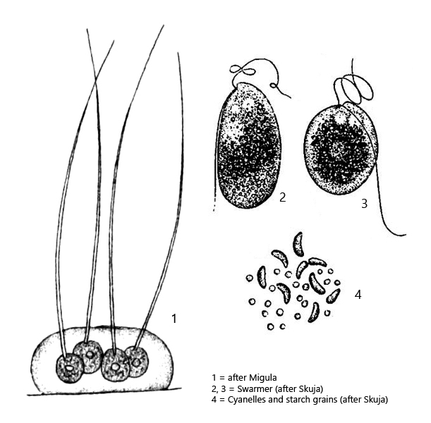
colonies of 2–4–8 spherical cells in a mucilagenous envelope
diameter cells 6–21 µm
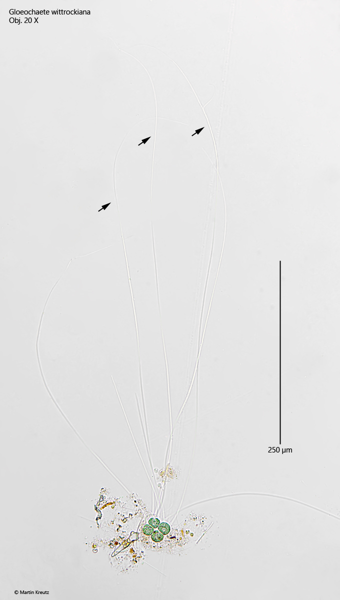
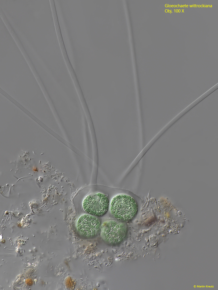
each cell with gelatinous setae, 100–500 µm long
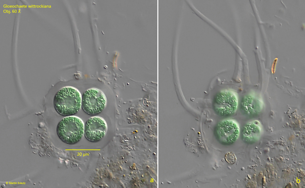
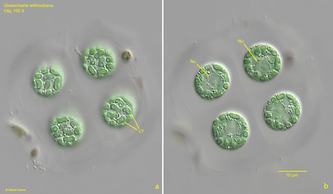
colored blueish-green by sausage-shaped cyanelles
cyanelles are arranged parietally
spherical nucleus central
Gloeochaete wittrockiana
So far I have only found Gloeochaete wittrockiana in the Simmelried, where the alga occurs rarely but regularly. In the samples, the colonies, which usually consist of 4 cells, are easily recognizable by their blueish-green color. The coloration is caused by so-called cyanelles, which are regarded as endosymbionts (s. fig. 4 a). In the course of evolution, cyanobacteria were taken up and integrated by the algae as symbionts. If an original chloroplast of the algae was present, it has been completely regressed.
Gloeochaete wittrockiana is classified as a tetrasporal alga because the cells can form very long gelatinous setae (s. fig. 1). In the literature their length is given as 100–250 µm, but I was also able to find colonies with bristles 500 µm long (s. fig. 1). However, they are very delicate and the thin ends can easily be overlooked.
The nucleus is located in the center of the cell. It appears to lie in a separate vacuole, which is free of cyanelles and which is separated from the ectoplasm by a granular layer (s. fig. 4 b).
Skuja (1956) described that Gloeochaete wittrockiana can form swarmer with two flagella and two contractile vacuoles (s. drawings above). I myself have not yet been able to observe such swarmers.
Fig. 1:Gloeochaete wittrockiana. L = appr. 500 µm (of colony with gelatinous setae). A colony of 4 cells with long, delicate setae (arrows). The setae have a length of about 500 µm. Obj. 20 X.
Fig. 2 a-b:Gloeochaete wittrockiana. D = 13–15 µm (of cells). Two focal planes of a colony of 4 cells. Obj. 60 X.
Fig. 3:Gloeochaete wittrockiana. D = 15–17 µm (of cells). A colony of 4 cells in a mucilagenous envelope. Obj. 100 X.
Fig. 4 a-b:Gloeochaete wittrockiana. D = 12–13 µm (of cells). Focal plane on the blueish-green cyanelles (CY, a) and the nuclei (Nu, b). The cyanelles have a length of 3.5 µm. Obj. 100 X.
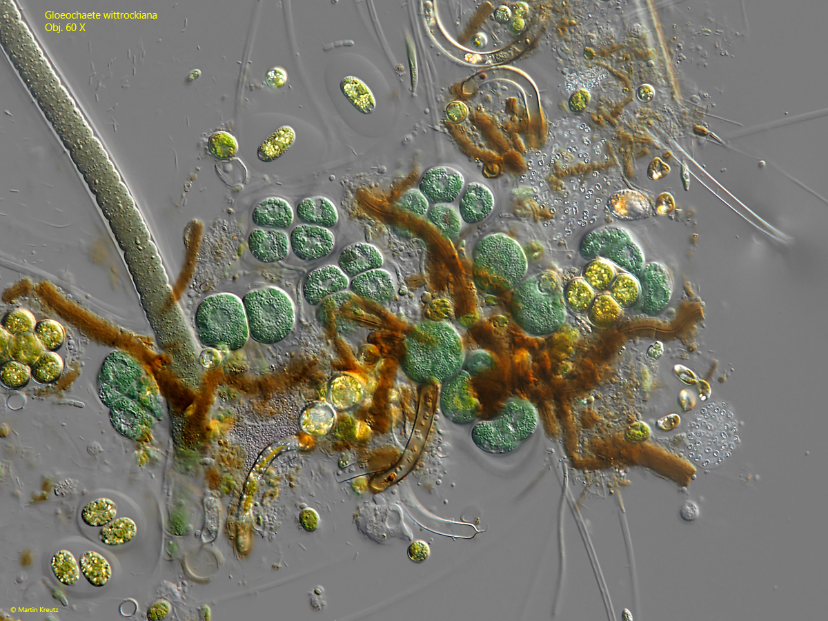
Fig. 5:Gloeochaete wittrockiana. A cluster of cells, mixed up with different species of algae, cyanobacteria and purple bacteria. Obj. 60 X.