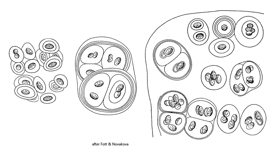
colonies irregularly shaped, amorphous and mucilaginous
cells irregularly distributed in the colonies
one, two or four cells in concentrically layered envelopes
one chloroplast, cup-shaped, filling two-thirds of cell
older cells filled with oil droplets and starch grains
one pyrenoid
Gloeocystis polydermatica
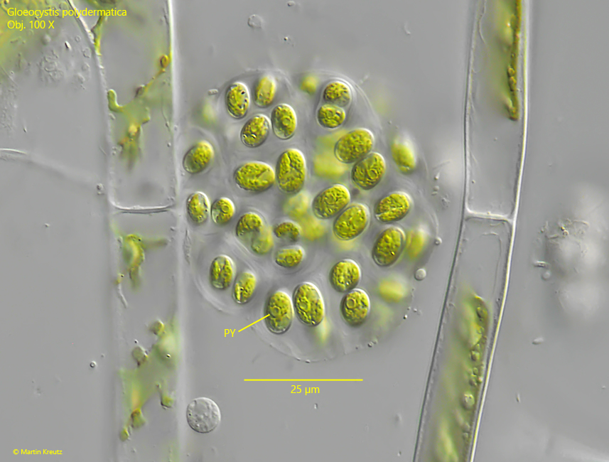
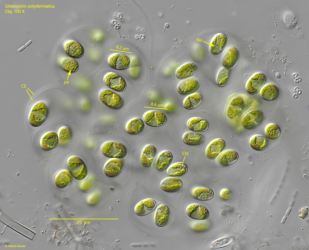
I find Gloeocystis polydermatica regularly but rarely in the Simmelried. The colonies should be about 20 X 30 µm according to Hindák (1978). This is also about the size of the colonies in my population with diameters of 20–70 µm (not squashed). The cells in my population were mostly between 8–10 µm long and oval. The chloroplast is cup-shaped (s. fig. 4) and I could observe one pyrenoid (s. figs. 1 and 4). The cells were irregularly distributed in the colony and surrounded by a concentrically layered envelope (s. figs. 3 and 4). Thus, all features agree with the description of Hindák.
Fig. 1:Gloeocystis polydermatica. D = 52 µm (of colony). A colony of about 50 cells. Obj. 100 X.
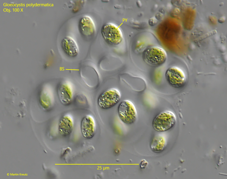
Fig. 2:Gloeocystis polydermatica. A slightly squashed, small colony with some blank spaces (BS). Obj. 100 X.
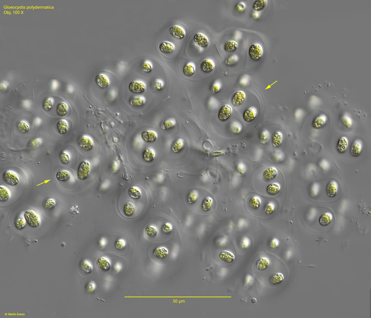
Fig. 3:Gloeocystis polydermatica. A slightly squashed larger colony. Note the concentracally layered envelope covering the cells (arrows). Obj. 100 X.
Fig. 4:Gloeocystis polydermatica. L = 8.0 – 9.1 µm (of cells). A slightly squashed colony in detail. Note the cup-shaped chlorplast (Chl) of the cells. CE = concentrically layered envelope, Nu = nucleus, PY = pyrenoid. Obj. 100 X.