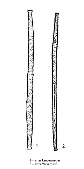
cell long, spindle shaped, tapering to slightly swollen capitate apices
cell wall covered with granules or short conical spines
length 150–250 µm, width 7–11 µm
two ribbon-shaped chloroplasts with 5–16 pyrenoids
chlorplast fill not the capitate apices
nucleus central
Gonatozygon brebissonii
I find Gonatozygon brebissoni rarely, but regularly. The specimens are usually found in shallow places in the sampling sites where they are found in the uppermost mud layer.
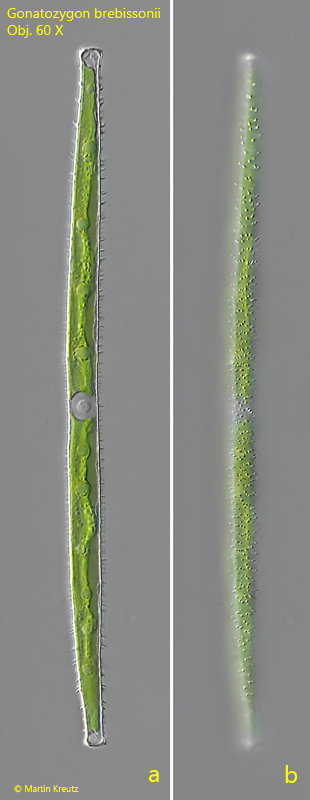
Gonatozygon brebissonii can be identified even at small magnifications, as the apical ends are clearly swollen. The cells are straight and at most minimally curved. The most striking feature is the clearly granulated or with short spines covered cell wall. In my population there are short spines (s. fig. 1 b and 2 b). The apical ends are not filled with the chloroplasts and remain transparent. However, there are no vacuoles filled with crystals as in Closterium.
Fig. 1 a-b:Gonatozygon brebissonii. L = 172 µm. Two focal planes of a specimen covered with short spines. Obj. 60 X.
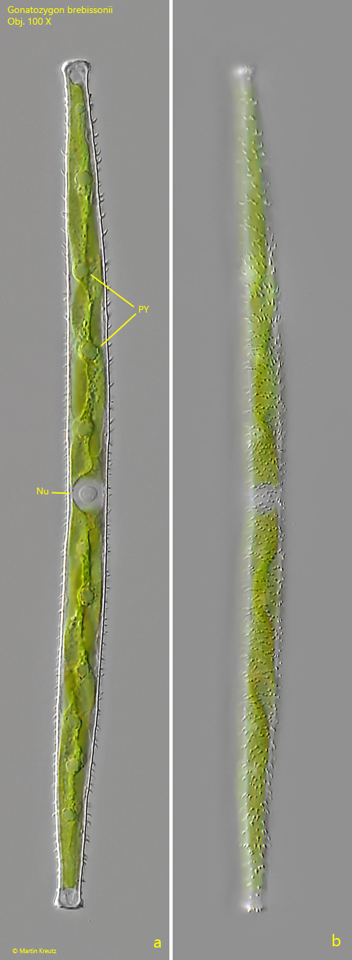
Fig. 2 a-b:Gonatozygon brebissonii. L = 172 µm. The same specimen as shown in fig. 1 a-b in detail. Nu = nucleus, PY = pyrenoids. Obj. 100 X.