end of cells transparent, often with vacuoles containing some crystals
spherical nucleus centrally between the chloroplasts
Gonatozygon kinahanii
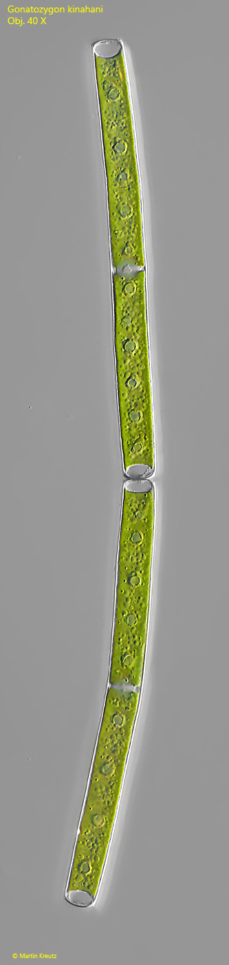
I find Gonatozygon kinahanii regularly, but rarely in the Simmelried. Up to now I have found exclusively single cells and a pair of cells (s. fig. 5). Filaments with several cells I have not found yet.
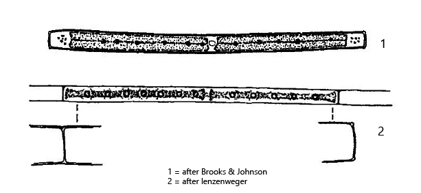
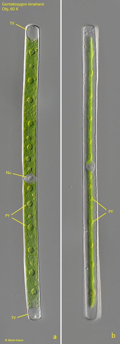
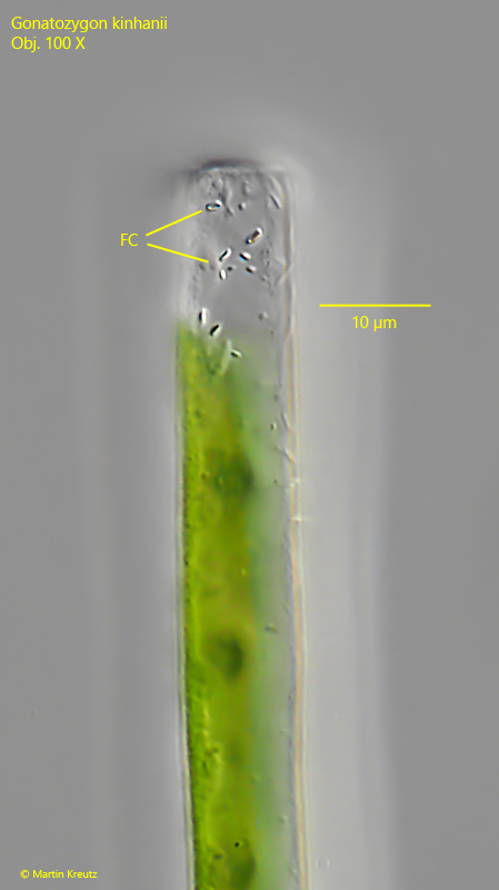
Gonatozygon kinahanii can be identified by the shape of the cell ends which neither widen nor taper. The cell ends are transversely truncated. Sometimes the cell ends are still convexly rounded (s. fig. 1 a). The two chloroplasts are symmetrically aligned in both halves of the cell. Their flat and ribbon-like shape can be seen by carefully rotating the cell under the coverslip (s. fig. 1 a-b). In the cytoplasm, there are often small, colorless crystals, which cluster especially in the terminal vacuoles (s. figs 4 and 5).
Fig. 1 a-b:Gonatozygon kinahanii. L = 266 µm. Focus on the broad side of the ribbon-shaped chloroplasts (a) and on the narrow edge of the chloroplasts after turning of the cell by 90° (b). Nu = nucleus, PY = pyrenoids, TV = terminal vacuoles. Obj. 60 X.
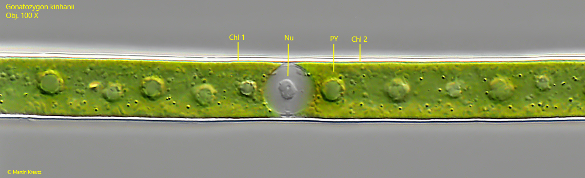
Fig. 2:Gonatozygon kinahanii. Detail of the cell shown in fig. 1 a-b. The nucleus (Nu) is located in the middle between the two chloroplasts (Chl 1, Chl 2). PY = pyrenoids. Obj. 100 X.
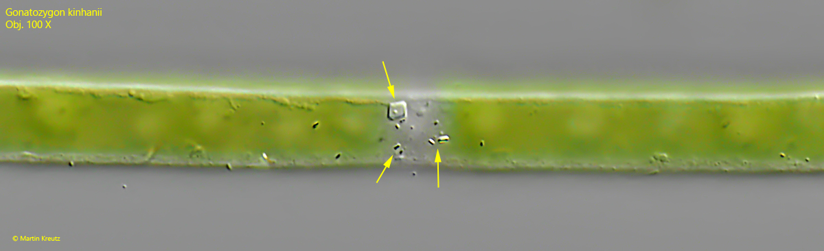
Fig. 3:Gonatozygon kinahanii. Focal plane on the crystals floating in the cytoplasm of the cell (arrows). Some of them have the shape of square tiles. Obj. 100 X.
Fig. 4:Gonatozygon kinahanii. Focal plane on the floating crystals (FC) in one of the terminal vacuoles of the cell. Obj. 100 X.
Fig. 5:Gonatozygon kinahanii. 228 and 236 µm. A pair of cells, probably after a cell division. Obj. 40 X.