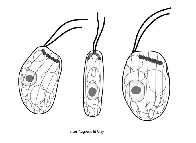
four to six ejectisomes arranged in parallel to anterior margin
one contractile vacuole opposite of flagella
periplast with 2–4 slightly oblique stripes, sometimes absent
nucleus in mid-body below position of flagella
Goniomonas truncata
Goniomonas truncata is a very common flagellate that belongs to the Cryptomonads. Unlike other Cryptomonads such as Cryptomonas or Rhodomonas, Goniomonas has no chloroplasts and is therefore colorless. In addition, the ejectisomes do not line the pharynx, as in Cryptomonas, for example, but are arranged in a parallel row at the anterior end (s. fig. 3c). Although Goniomonas truncata was first described about 150 years ago, the fine striation of the periplast was not discovered until 1939 by Skuja. Sometimes this striation is missing. However, in my populations all specimens showed this striation (s. figs. 2c, 3d, 3e and 4c). On each side 3–4 stripes can be seen. In addition, I could see a distinct punctation of the stripes, which to my knowledge has not been described yet.
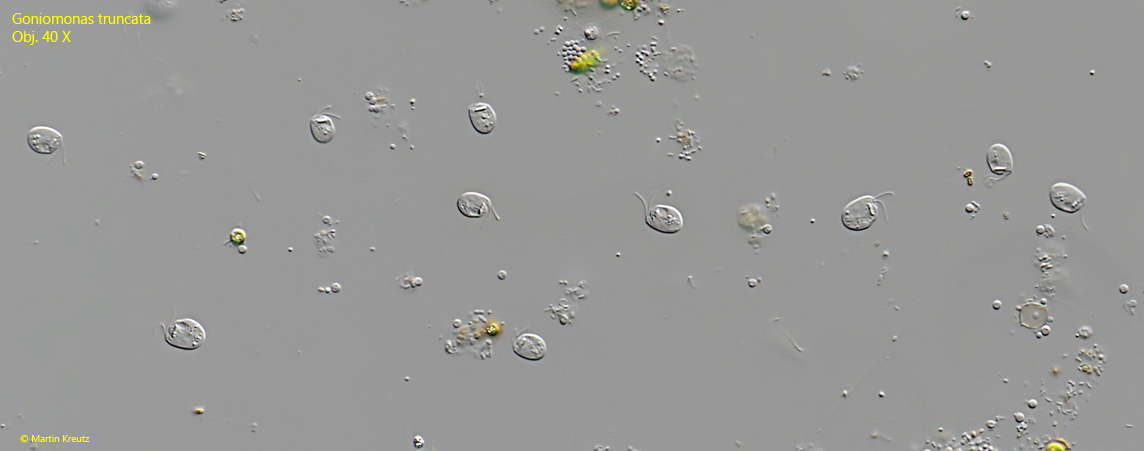
Goniomonas truncata is easy to observe, because the flagellates like to accumulate on floating coverslips, which are placed on the surface of the samples. After a few days, there may be hundreds of specimens depending on the underlying samples (s. fig. 1). Samples with decaying plant matter are best.
Fig. 1:Goniomonas truncata. Overview of an assemblage of specimens that have gathered on a floating coverslip. Obj. 40 X.
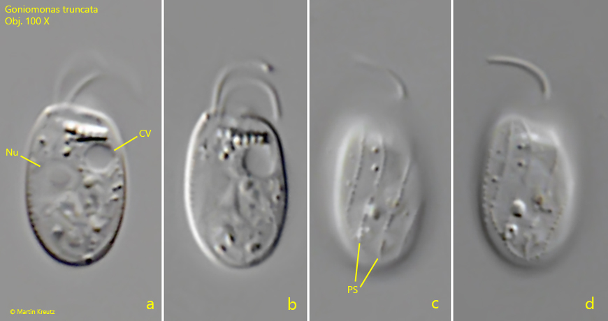
Fig. 2 a-d:Goniomonas truncata. L = 9.8 µm. Different focal planes of a freely swimming specimen. Note the delicate striation of the periplast (PS). CV = contractile vacuole, Nu = nucleus. Obj. 100 X.
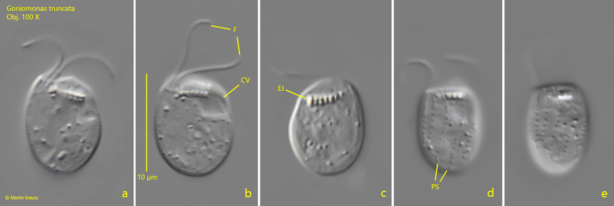
Fig. 3 a-e:Goniomonas truncata. L = 10.0 µm. Different focal planes of a second freely swimming specimen. Note the two flagella (F) of almost equal length and the ejectisomes (EJ) arranged in a parallel row near the anterior margin. CV = contractile vacuole, PS = striation of the periplast. Obj. 100 X.
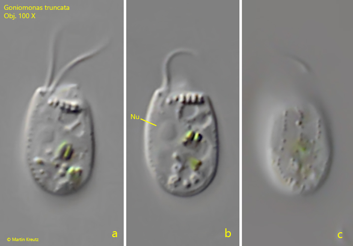
Fig. 4 a-c:Goniomonas truncata. L = 9.7 µm. Different focal planes of a third freely swimming specimen. The nucleus (Nu) is located near mid-body on the the side where the flagella emerge. Obj. 100 X.