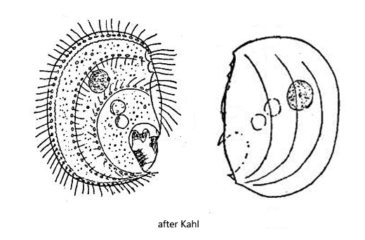
cytostome in depression on the posterior ventral edge, depression opens more to the ventral side
tooth-like structure on the anterior edge of the oral depression
two contractile vacuoles above the oral opening
macronucleus spherical in mid-body
one spherical micronucleus
Hemicyclium lucidum
I have found Hemicyclium lucidum so far exclusively in the uppermost mud layer in Simmelried, but there the species occurs regularly. In some samples more than 10 specimens per milliliter are present. The species is very easy to recognize by the 3 well visible rows of cilia on the ventral side and because the cilia arise in pairs from distinct pores (s. figs. 2 a-b, 3 and 4). Compared to similar species of the genus Microthorax, Hemicyclium lucidum is much larger and the mouth opening is not located at the end of the body but in the posterior third. In addition, Hemicyclium lucidum has a distinct tooth or spine above the mouth opening (s. fig. 2b).
Upon close examination of the ventral side of the body, in addition to the three rows of pores from which the cilia arise, I was able to note another pore-like ornamentation of the pellicle (s. fig. 3). These pores are less strongly outlined and therefore more inconspicuous. Also, they are not perfectly round, but slightly deformed. To my knowledge, this ornamentation has not been observed by previous authors.
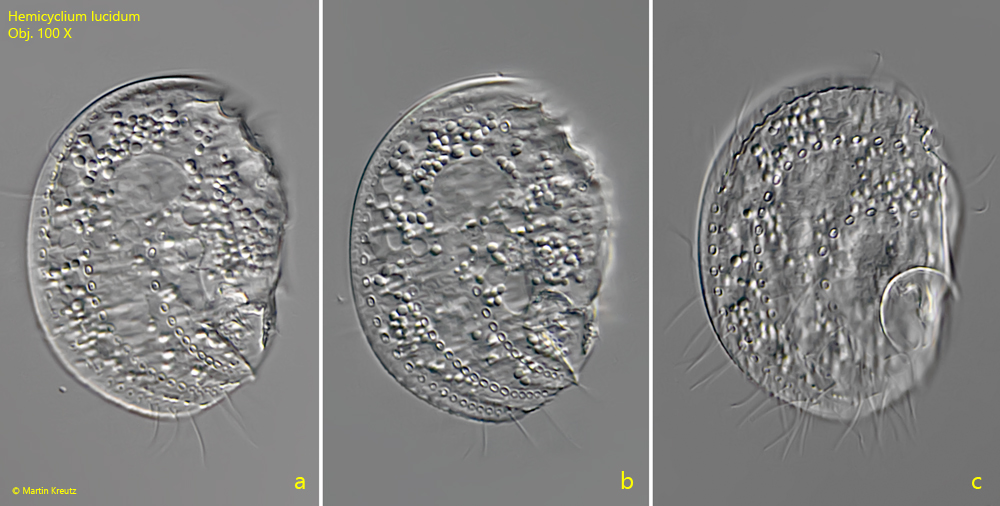
Fig. 1 a-c:Hemicyclium lucidum. L = 62 µm. Ventral view of a freely swimming specimen. Obj. 100 X.
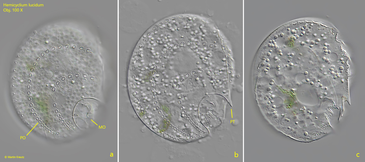
Fig. 2 a-c:Hemicyclium lucidum. L = 55 µm. Three focal planes of the ventral side of a second specimen. MO = mouth opening, PO = pores, PT = pre-oral tooth. Obj. 100 X.
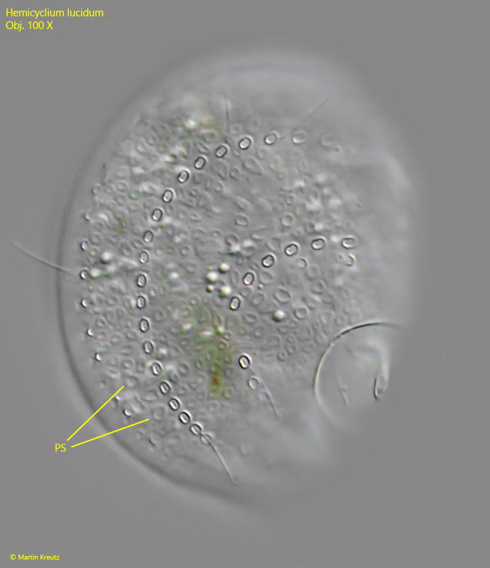
Fig. 3:Hemicyclium lucidum. L = 55 µm. The same specimen as shown in fig. 2 a-c with a slightly lower focus on the pellicle. A pore system (PS) becomes visible. These pores are all circular but different in shape and size. Obj. 100 X.
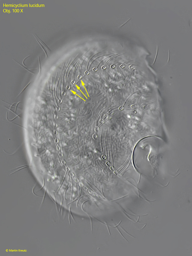
Fig. 4:Hemicyclium lucidum. L = 71 µm. Focus on the ventral side of a slightly squashed specimen. Two cilia arise from each pore (arrows). Obj. 100 X.
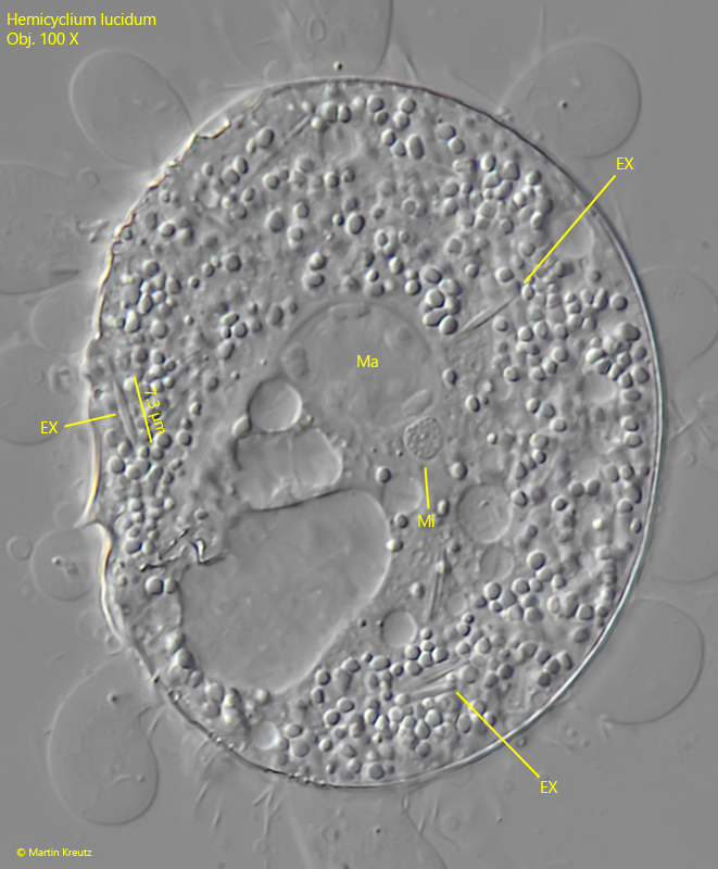
Fig. 5:Hemicyclium lucidum. In a strongly squashed specimen the macronucleus (Ma) and micronucleus (MI) are visbles as well the spindle-shaped extrusomes (EX) with a length of 7–8 µm. Obj. 100 X.
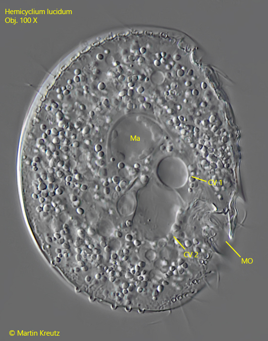
Fig. 6:Hemicyclium lucidum. Above the mouth opening two contractile vacuoles are located (CV 1, CV 2). Due to the pressure of the coverslip CV 2 is enlarged. Obj. 100 X.