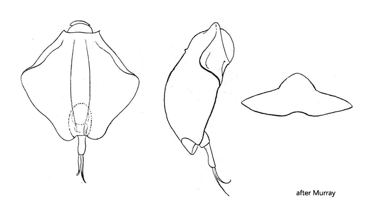
terminal segment of foot longer than basal foot section
pointed toes of different lengths
Heterolepadella heterostyla
I found Heterolepadella heterostyla in the Spechtensee in Styria/Austria in June 1999 and 25 years later in June 2024 in the Sima Moor (Austria). This species can easily be confused with the rhombic form variant of Lepadella triptera. However, Heterolepadella heterostyla has toes of different lengths (s. figs. 1 b, 2 a and 5). The right toe is always the shorter one. The lorica has a rhombic shape and the lateral corners are slightly bent forward (s. fig. 2 a). A triangular-shaped rostrum can only be seen in an outstretched specimen (s. fig. 2 b). On the ventral side of the lorica of specimens from the Sima Moor I could recognize 7 humps, which are not described in the literature (s. fig. 3).
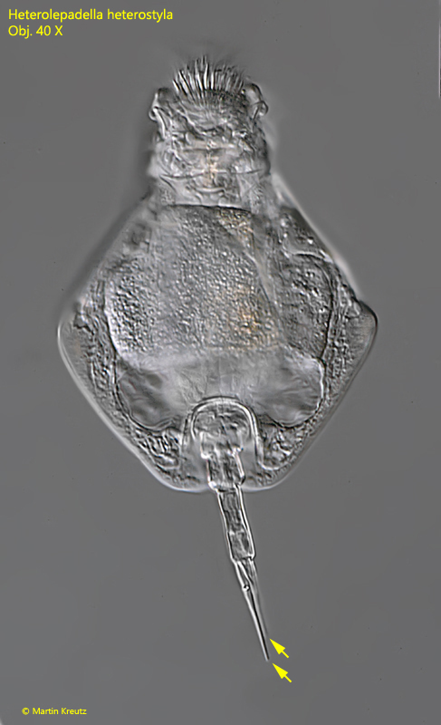
Fig. 1 a-d:Heterolepadella heterostyla. L = 85 µm (of the lorica). A slightly squashed specimen from ventral. Obj. 40 X.
Fig. 2 a-b:Heterolepadella heterostyla. L = 85 µm (of the lorica). The slightly squashed specimen as shown in fig. 1 a-d in detail. Note the different lengths of the toes (TO) and the granulated collar (GC). RO = rostrum. Obj. 60 X.
Fig. 3:Heterolepadella heterostyla. L = 85 µm (of the lorica). Focal plane on the 7 humps (1-7) visible on the ventral side of the lorica. Obj. 60 X.
Fig. 4:Heterolepadella heterostyla. L = 82 µm (of the lorica). Ventral view of a slightly squashed specimen found in June 1999 in the Spectensee (Austria). Obj. 40 X.
Fig. 5:Heterolepadella heterostyla. The three segments of the foot (FT) and the toes (TO) of different length in a squashed specimen. Obj. 100 X.
Fig. 6 a-b:Heterolepadella heterostyla. Two focal planes of the trophi in a strongly squashed specimen. Obj. 100 X.