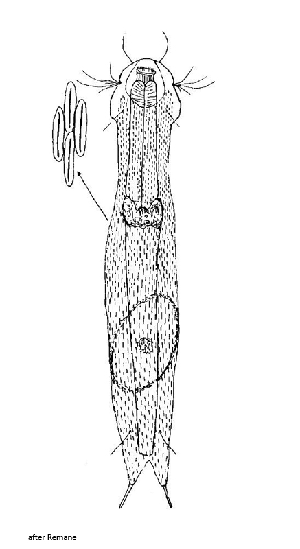
body slender, elongate, L = 130–200 µm, W = 29–30 µm
head trilobed, 21–23 µm wide
two lateral ciliary tufts
anterior ciliary tufts only consisting of one sensory hair
cephalion large, covering the head widely
posterior pleural lobes well developed
hypostomium developed as 2 cusps
neck distinctly constricted, elongate, on average 18-19 µm wide
two dorsal setolae on strongly incised special scales
furca 14–20 µm long, base of toes fully scaled
free adhesive tubes occupying half of toe length (7–11 µm)
approximately 25 dorsal longitudinal rows, each with 40–80 elongate narrow keel scales
ventral field with 2 terminal keel plates, otherwise 6–11 longitudinal rows, each with 18–30 small keel scales (1.5–3 µm)
pharynx cylindrical, terminally little swollen, 35–51 µm long
anterior end of intestine delimited with a golden brown ring
Heterolepidoderma majus
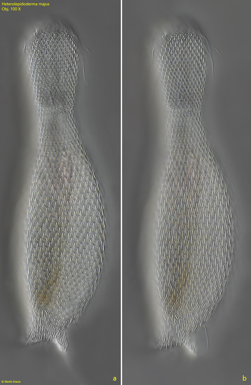
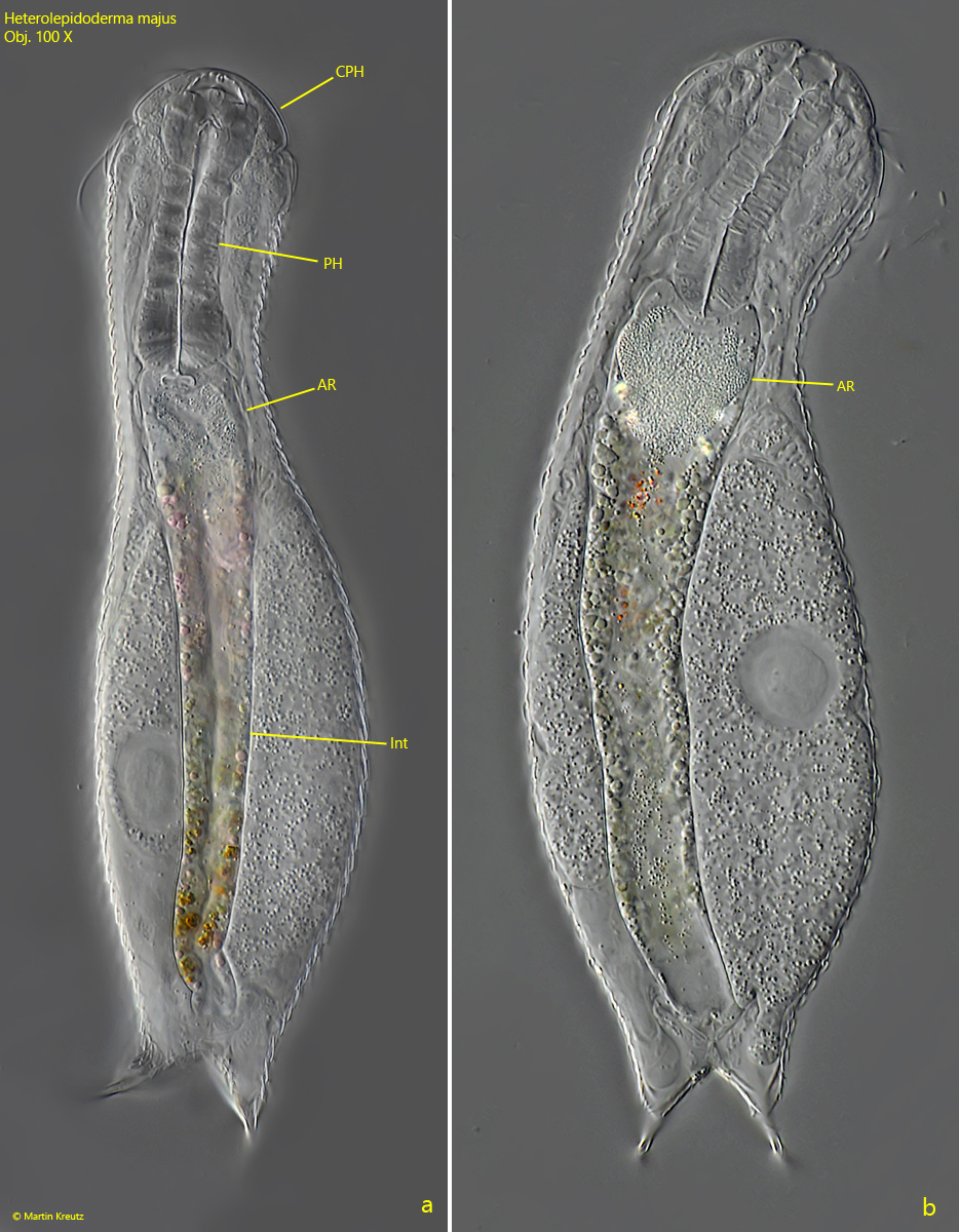
I have found Heterolepidoderma majus in June 2022 in Simmelried in decomposing plant material. The exact shape of the keeled, elongate scales is difficult to recognize in the living specimen. Depending on the focus, it may appear that the scales are round (s. fig. 1a). However, this is due to the fact that the scales lie in a slightly oblique, roof tile-like way on top of each other. In optical section, this can give the impression of a round shape. However, the long, somewhat protruding keels are clearly visible. The “golden brown” ring, which lies at the anterior end of the intestine (s. fig. 2), usually appears colorless or yellowish in DIC. Only in brightfield illumination the yellow-brownish coloration can be seen.
Fig. 1 a-b: Heterolepidoderma majus. L = 170 µm. Two focal planes of the dorsal scales of a slightly squashed specimen. Obj. 100 X.
Fig. 2 a-b: Heterolepidoderma majus. L = 170 µm. Focal plane on the intestine (Int) of two separate specimen (a, b). AR = anterior ring of intestine, CPH = cephalion, PH = pharynx. Obj. 100 X.
Fig. 3: Heterolepidoderma majus. L = 154 µm. Focal plane on the dorsal scales of a squashed specimen. Obj. 100 X.
Fig. 4: Heterolepidoderma majus. The dorsal scales in mid-body in detail. Obj. 100 X.
Fig. 5: Heterolepidoderma majus. The dorsal scales at the posterior end in detail. Obj. 100 X.
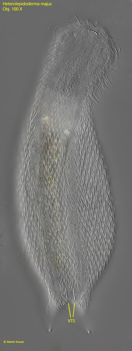
Fig. 6: Heterolepidoderma majus. L = 185 µm. A squashed specimen in ventral view. VTS = ventral terminal scales. Obj. 100 X.
Fig. 7: Heterolepidoderma majus. The ventral scales (VS) in detail. VTS = ventral terminal scales. Obj. 100 X.