trailing flagellum 1/4 length of locomotion flagellum
Heteronema mutabile
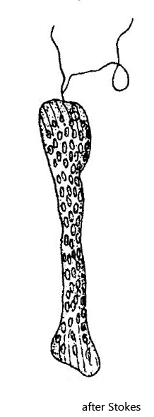
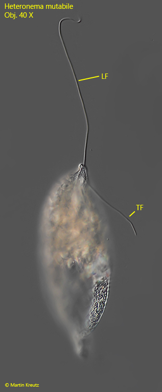
In my sampling site Simmelried I regularly and frequently find a very large flagellate with a locomotion flagellum and a trailing flagellum. Most specimens are 150–200 µm long and always completely filled with highly refractive paramylon grains. Based on the two flagella and the round body shape in cross section it is a member of the genus Heteronema. Within the genus Heteronema only the species Heteronema mutabile reaches a body length of more than 150 µm (up to a maximum of 253 µm according Stokes). The cells are very metabolic, with a distinct striation of the pellicle (s. fig. 3 a-b). The locomotion flagellum reaches body length, as described by Stokes (s. fig. 2). Usually the cells are completely opaque due to highly refractive paramylon granules or phagocytosed algae (s. figs. 1 a-b, 3 a-b and 6 a-c). The nucleus is sub-spherically shaped with a large central nucleolus (s. fig. 4 b). At the anterior end, near the reservoir, the rods of the ingestion apparatus are visible (s. fig. 6 a-c). With the help of these rods the mouth opening is widened and the flagella are shifted laterally, so that the prey can pass the gullet.
The description and the drawing of Stokes are not very informative. But I have not found any further descriptions of this species. However, because of the body size of this Heteronema species there are no alternatives and since all the characteristics described by Stokes are indeed present, I believe that my findings represent Heteronema mutablile as well. Yet the specimens of my population also show great similarity with Jenningsia diatomophaga. This species is supposed to grow even larger than Heteronema mutabile. Indeed, some specimens of my population were 290 µm long (s. fig. 4a-c). However, Jenningsia is said to have only one flagellum and feeds exclusively on diatoms. The descriptions of Jenningsia diatomophaga available to me were obviously made on freely moving specimens. Therefore, I do not consider it impossible that the trailing flagellum, which is closely attached to the body, may have been overlooked. The specimens in my population feed mainly on algae and dinoflagellates. Whether Jenningsia diatomophaga is really a food specialist remains questionable based on the few observations. Therefore, only further investigations can clarify whether Heteronema mutabile and Jenningsia diatomophaga are possibly the same species.
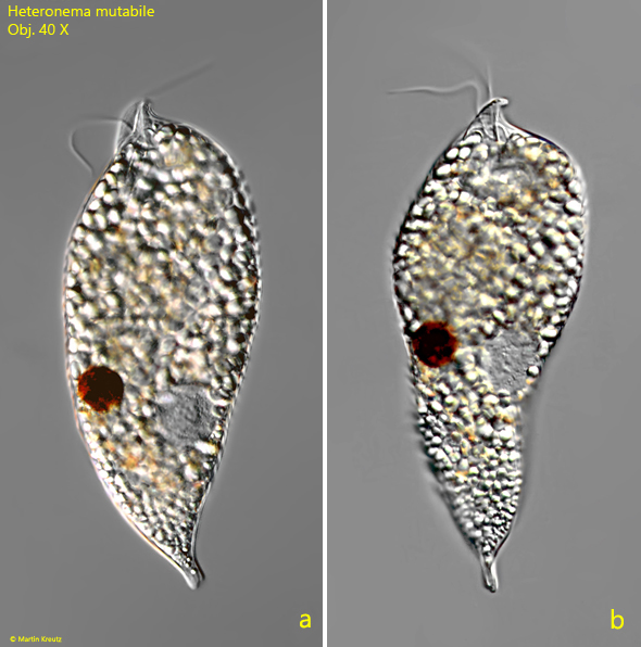
Fig. 1 a-b: Heteronema mutabile. L = 196 µm. A slightly squashed specimen. Note the funnel-shaped apical dilation of the cytospome (“pharynx”). Obj. 40 X.
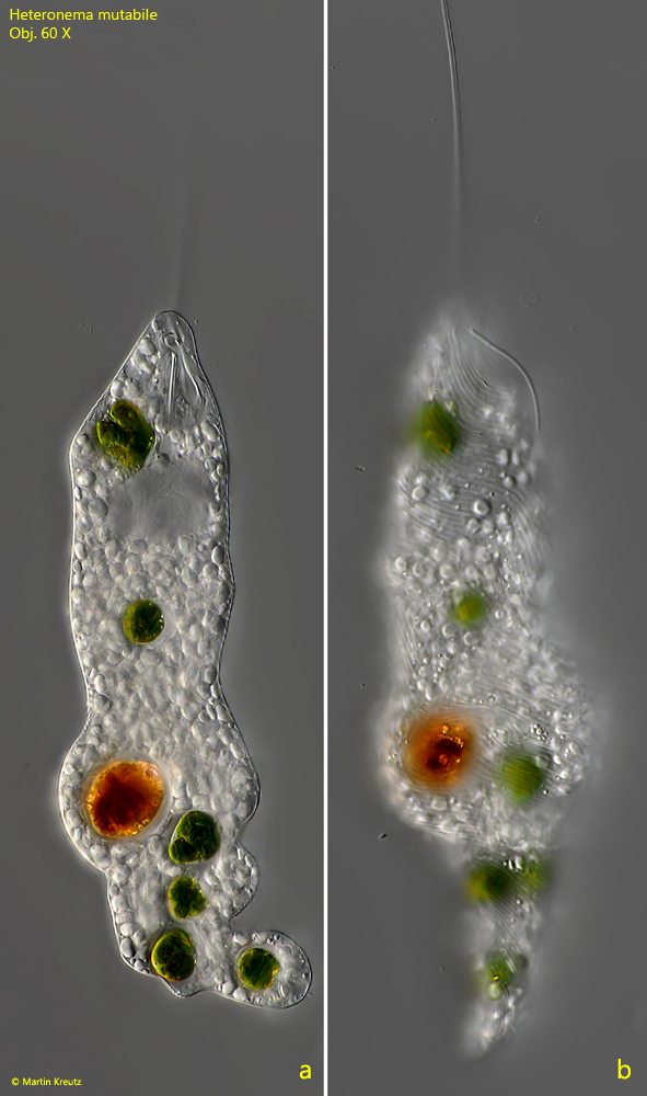
Fig. 3 a-b: Heteronema mutabile. L = 205 µm. Two focal planes of a slightly squashed specimen. Note the striation of the pellicle (b). Obj. 60 X.
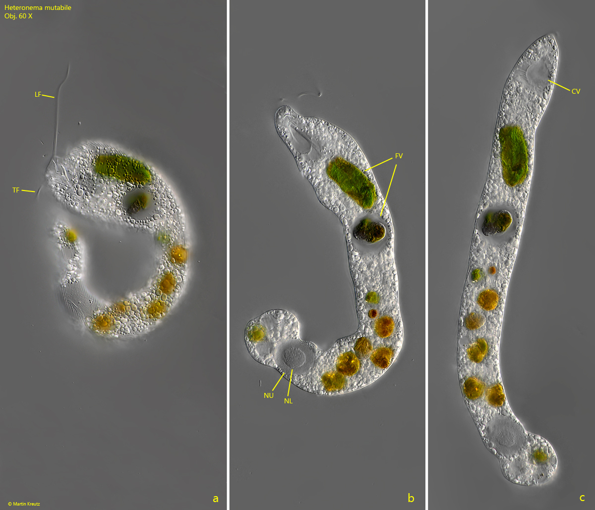
Fig. 4 a-c: Heteronema mutabile. L = 290 µm. The metabolic movenment of a second specimen. CV = contractile vacuole, FV = food vacuoles, LF = locomotion flagellum, NL = nucleolus, Nu = nucleus, TF = trailing flagellum. Obj. 60 X.
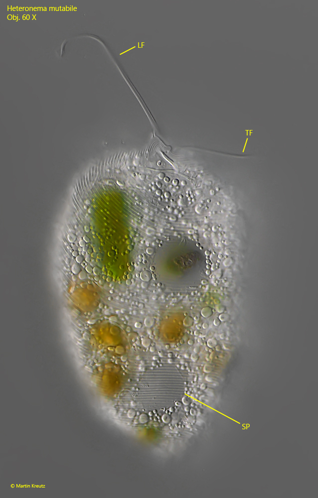
Fig. 5: Heteronema mutabile. Focus on the striation of the pellicle (SP) of a contracted specimen. LF = locomotion flagellum, TF = trailing flagellum. Obj. 60 X.
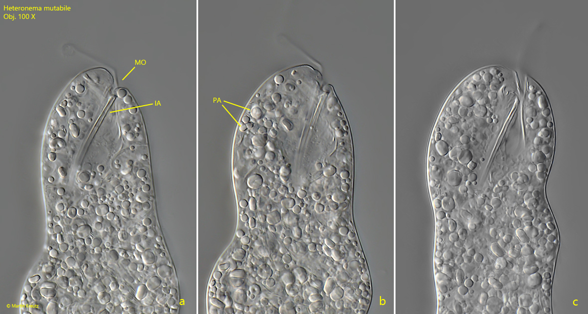
Fig. 6 a-c: Heteronema mutabile. Three focal planes of the anterior end of a specimen with the clearly visible rods of the ingestion apparatus (IA) and the mouth opening. MO = mouth opening, PA = paramylon bodies. Obj. 100 X.
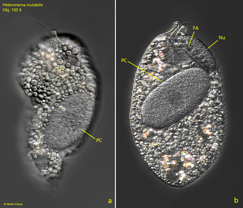
In my population of Heteronema mutabile there were many specimens infected by a parasitic fungus. In the infested specimens, a 40–60 µm long, oval-shaped cell of the fungus could be seen in the plasma (s. fig. 7 a-b). In most cases, this fungal cell was centrally located in the middle of the body. The nucleus of the host cell was shifted towards the mouth opening or the posterior end. I could observe a similar infestation also in other Euglenophyceae of the genera Phacus, Lepocinclis and Euglena. I could not follow the life cycle of this parasitic fungus.
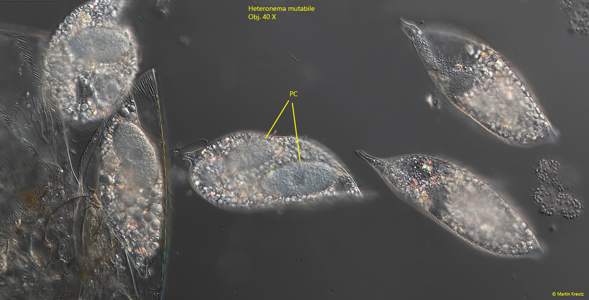
Fig. 7 a-b: Heteronema mutabile. L = 150 µm. A specimen infested with a parasitic fungus cell (PC). FA = feeding apparatus (= ingestion apparatus), Nu = nucleus. Obj. 100 X.
Fig. 8: Heteronema mutabile. L = 150–160 µm. Some specimens feeding on a dead crustacean. Three of these specimens are infested by a parasitic fungus. The large parasitic fungi cells (PC) are visible inside the cells. The labeled cell is infected by even two parasitic cells. Obj. 100 X.