body spindle-shaped, spirally twisted usually with 3 turns
anterior and posterior ends are extended teat-shaped
length 96–130 µm, width 39–46 µm
leading flagellum about body length
trailing flagellum about half as long as body
periplast with spirally striation
nucleus central
pharynx with rod-shaped organell
body filled with numerous roundish paramylon grains
Heteronema trispira
I find Heteronema trispira comparatively rarely. So far, all specimens have come from the uppermost mud layer in the Simmelried. In rare cases, Heteronema trispira also settles on the floating coverslip.
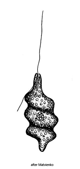
Only the origninal description and drawing of Heteronema trispira by Matvienko (1938), who found this Heteronema species in a Russian Sphagnum pond, seems to exist. After that there seem to be no further records of this species.
Heteronema trispira is slender spindle-shaped and has three characteristic coils which are turned counterclockwise. The anterior end is snout-like and the posterior end is tapered and rounded, as described and drawn by Matvienko. In my population, however, there are some deviations from Matvienko’s description. In my specimens, the leading (swimming) flagellum was only about half as long as the body and the trailing flagellum only slightly shorter. Matvienk describes them as body-length. I could not find the rod-shaped organelle described by Matvienk in the area of the mouth opening in any of the specimens in my population.
The specimens in my population were 75–122 µm long, which fits well with Matvienk’s length data (96–133 µm). The smallest specimen with 75 µm also had three coils, but was completely filled with paramylon grains, which made the specimen appear rather clumsy (s. fig. 5 a-d). I was almost always able to identify phagocytized, small algae in the other specimens (s. fig. 3 c). Larger paramylon grains were often found in the anterior third, while clusters of very small paramylon grains were found in the posterior third. The nucleus was in the anterior third or in the center of the body. The striation of the pellicle can only be seen at high magnification between the the coils.
The similar species Heteronema spirale is only half the size (40–60 µm) and has 5–6 coils. This species is also plumper.
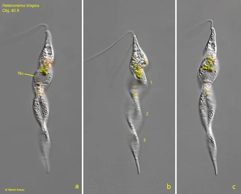
Fig. 1 a-c:Heteronema trispira. L = 122 µm. The freely swimming specimen found in January 1998. Note the three turns of the body (1–3). Nu = nucleus. Obj. 40 X.
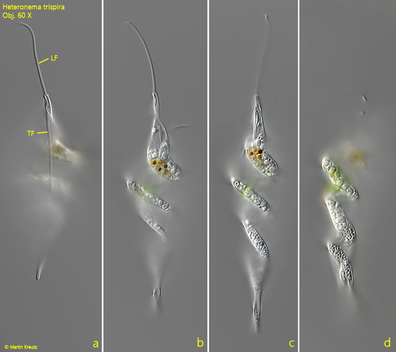
Fig. 2 a-d:Heteronema trispira. L = 112 µm. Different focal planes of a second specimen. Note the leading flagellum (LF) and the trailing flagellum (TF). Obj. 60 X.
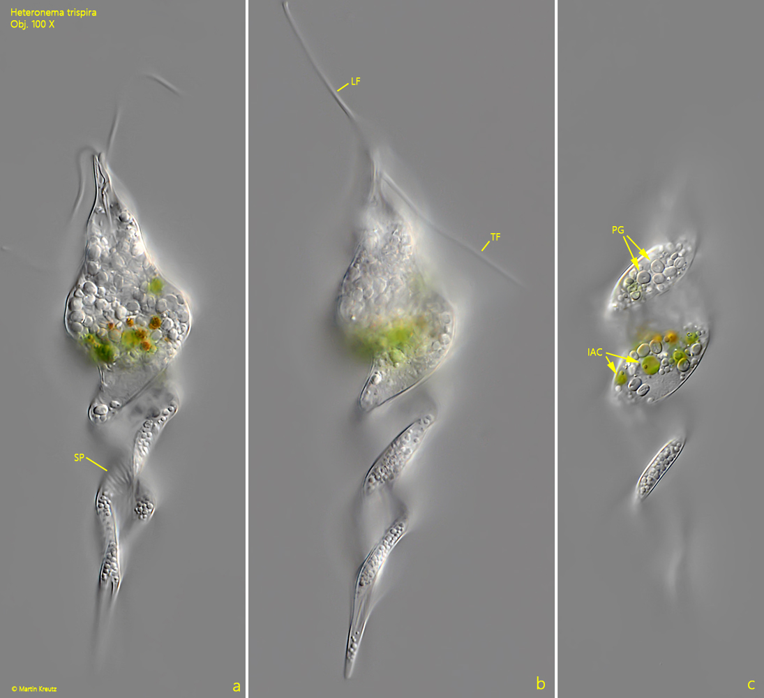
Fig. 3 a-c:Heteronema trispira. L = 112 µm. The slightly squashed specimen as shown in fig. 2 a-d at higher magnification. Note the striation of the pellicle (SP). IAC = ingested algae cells, LF = leading flagellum, PG = paramylon grains, TF = trailing flagellum. Obj. 100 X.
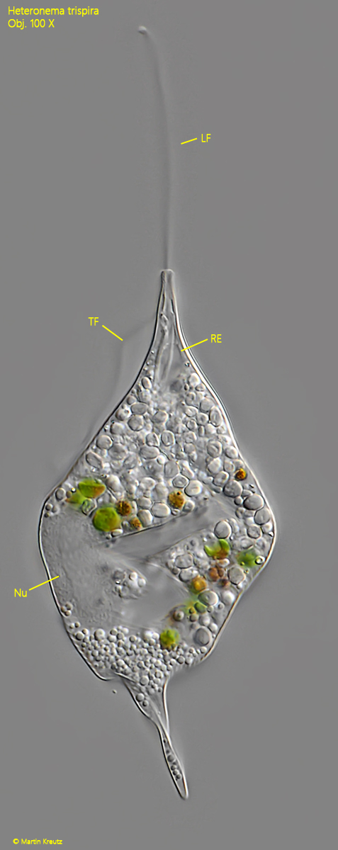
Fig. 4:Heteronema trispira. L = 112 µm. The squashed specimen as shown in fig. 2 a-d. LF = leading flagellum, Nu = nucleus, RE = reservoir, TF = trailing flagellum. Obj. 100 X.
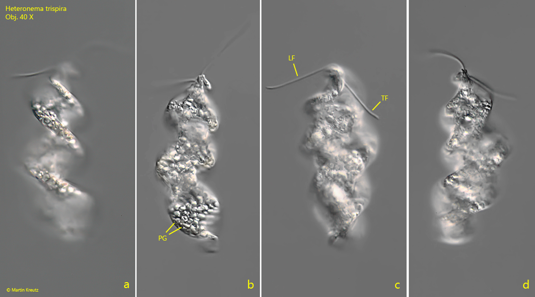
Fig. 5 a-d:Heteronema trispira. L = 75 µm. The third freely swimming specimen found in September 2003. LF = leading flagellum, PG = paramylon grains, TF = trailing flagellum. Obj. 40 X.
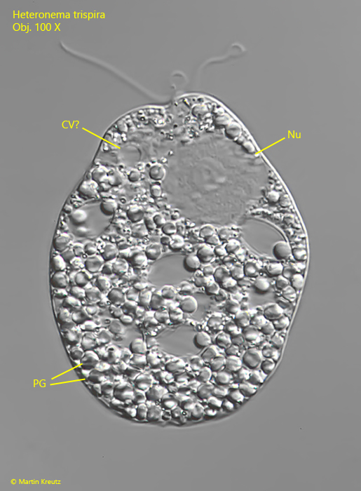
Fig. 6:Heteronema trispira. The strongly squashed specimen as shown in fig. 5 a-d. CV? = probably the contractile vacuole, Nu = nucleus, PG = paramylon grains. Obj. 100 X.