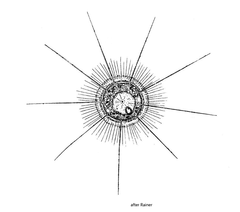
cell covered with thick mucous coat interspersed with randomly arranged, 10–13 µm long spicules
radially arranged spicules with the length of half the cell diameter penetrate the mucous coat
between cell and mucous coat a gap of about 5–10 µm is present
length of axopodia 1–2 cell diameter
single nucleus, located eccentrically
contractile vacuole usually absent
Heterophrys myriopoda
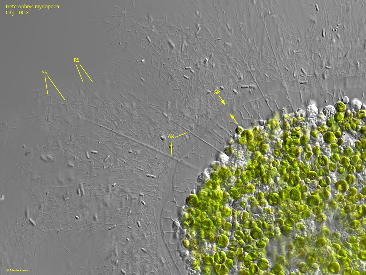
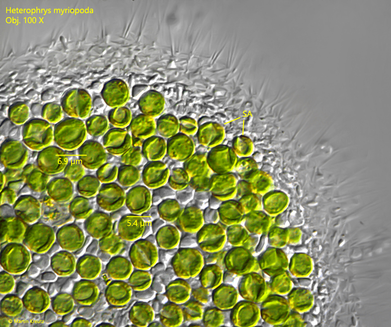
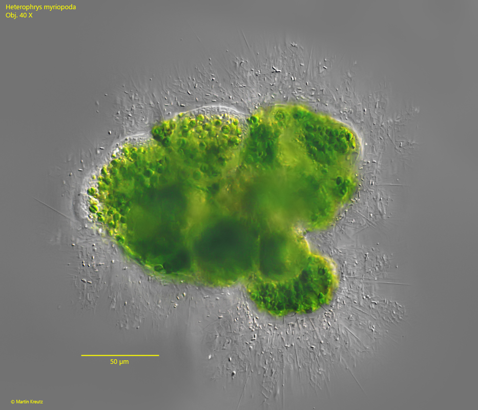
Heterophrys myriopoda was a very common heliozoan in Simmelried until about 2010. After that it became more and more rare. My last record is from July 2021. This heliozoan is easy to identify. First, the entire cell is filled with symbiotic algae and is therefore intensely green. The symbiotic algae appears to be of the Chlorella type with a diameter of 5–7 µm (s. fig. 6). The cell body is surrounded by a thick mucilaginous envelope in which short spicules are embedded and which often appears brownish in brightfield illumination. This mucilage envelope is pierced by very fine, radially extending spicules. In squashed specimens they are often not visible because they are “swallowed” by the squashed mucuos coat. Thicker and even longer are the axopodia, which radiate from the cell. Very characteristic in Heterophrys myriopoda is a small gap between the cytoplasm of the cell and the mucus coat (s. fig. 5). In one case I could observe a feeding community of Heterophrys myriopoda (s. fig. 7)
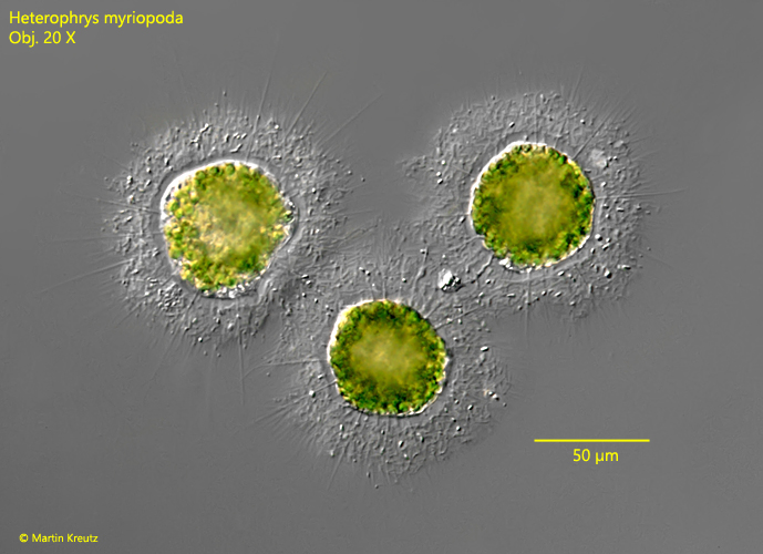
Fig. 1:Heterophrys myriopoda. D = 58 – 62 µm (without mucous coat). A group of three specimens. Obj. 20 X.
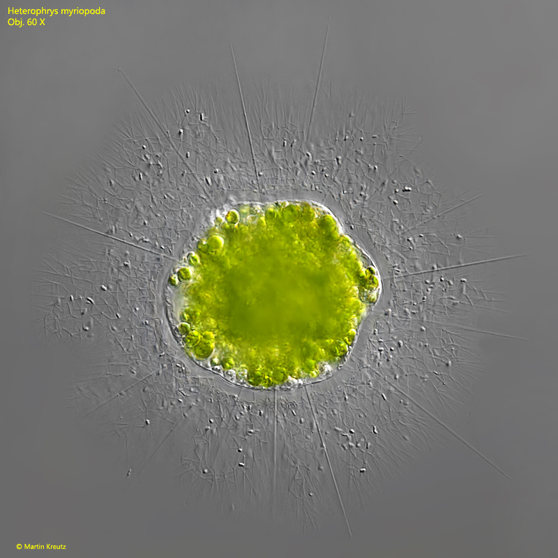
Fig. 2:Heterophrys myriopoda. D = 60 µm (without mucous coat). A slightly squashed specimen. AX = axopodia, MC = mucous coat with scattered spicules, RS = radial spicules. Obj. 60 X.
Fig. 3:Heterophrys myriopoda. A second slightly squashed specimen. Obj. 60 X.
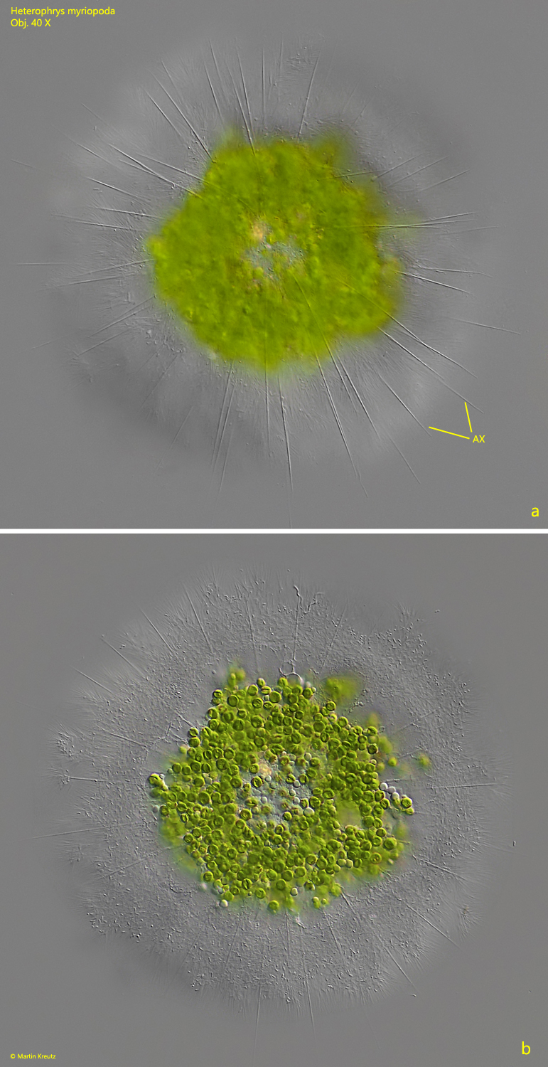
Fig. 4:Heterophrys myriopoda. Two focal planes of a slightly squashed specimen with focus on the axopodia (a, AX) and on the cell filled with symbiotic algae (b). Obj. 40 X.
Fig. 5:Heterophrys myriopoda. A strongly squashed specimen. Note the gap (GP, arrows) between the cytoplasm of the cell and the mucous coat. In the mucous coat thousands of short spicules (SS) are scattered. AX = axopodia, RS = radial spicules. Obj. 100 X.
Fig. 6:Heterophrys myriopoda. The symbiotic algae (SA) in a strongly squashed specimen. They have a diameter of 5–7 µm and appear to be of the Chlorella type. Obj. 100 X.
Fig. 7:Heterophrys myriopoda. A feeding community of about 10 individuals. They were partially merged, so that no single individuals could be clearly distinguished. Obj. 40 X.