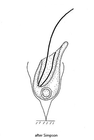
lorica is bell shaped, with a bulbous extension at level of the cell
cell with two flagella with different length
cell with a sail-shaped cytoplasmic lip
short flagellum moved or is attached to the cytoplasmic lip
spherical nucleus with central nucleolus in posterior third
contractile vacuole at the base of the cytoplasmic lip
Histiona aroides
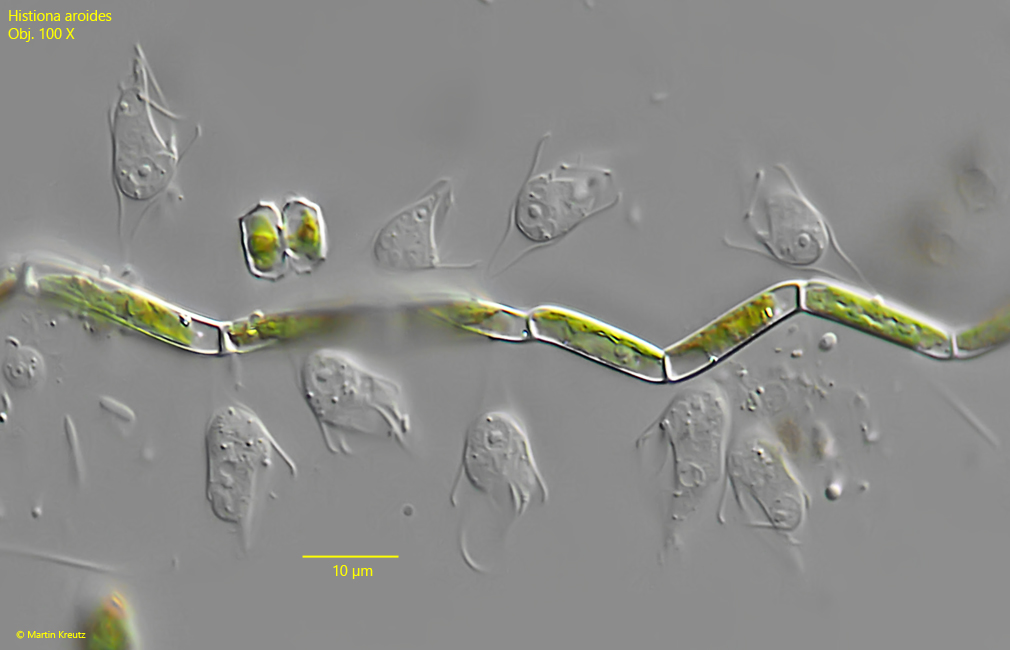
So far I could detect Histiona aroides only once in July 2021 in the Simmelried. I found colonies of 10 – 20 specimens each on algae filaments. The lorica of Histiona aroides has a campanulate shape, with a bulbous extension at the level of the cell. The flagellate in the lorica forms a sail-shaped cytoplasmic lip. Two flagella of different length arise from the base of this lip. The long flagellum swirls food (bacteria) in a whip-like manner. The function of the cytoplasmic lip seems to that of a funnel or the swirled bacteria stick to it and are then stripped off by the second, short flagellum. The short flagellum is difficult to see. It is supposed to be adjacent to the plasma lip. However, I could also observe that it moving over the plasma lip (s. fig. 3a-c).
Fig. 1:Histiona aroides. L = 15 -18 µm (from stalk of lorica to top of cytoplasmic lip). A colony on an alga filament. Obj. 100 X.
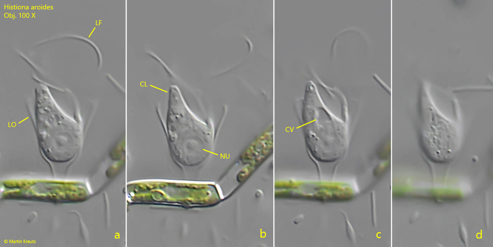
Fig. 2 a-d:Histiona aroides. L = 17 µm (from stalk of lorica to top of cytoplasmic lip). Different focal planes of a specimen. CL = sail-shaped cytoplamic lip, CV = contractile vacuole, LF = long flagellum, LO = lorica, Nu = nucleus with central nucleolus. Obj. 100 X.
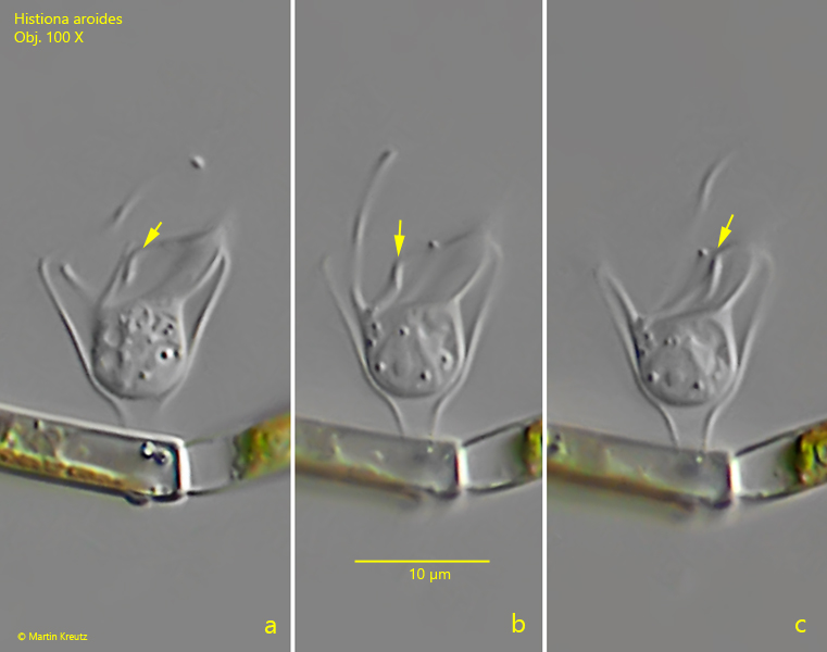
Fig. 3 a-c:Histiona aroides. L = 15 µm (from stalk of lorica to top of cytoplasmic lip). The short flagellum (arrow) is attached to the cytoplasmic lip or it moves over the lip. Obj. 100 X.