pharyngeal basket lined with thin, rod-shaped extrusomes
pharyngeal basket surrounded by large, comma- or spindle shaped extrusomes
pellicle coarse
macronucleus spherical or oval with one adjacent micronucleus
contractile vacuole terminal
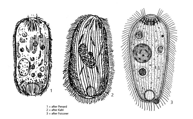
Holophrya saginata
So far I have found only two specimens of Holophrya saginata. The first specimen in August 2007 in the Simmelried and the second specimen in September 2023 in the Ulmisried. The rare finds are probably due to the fact that Holophrya saginata is actually known as a moss form.
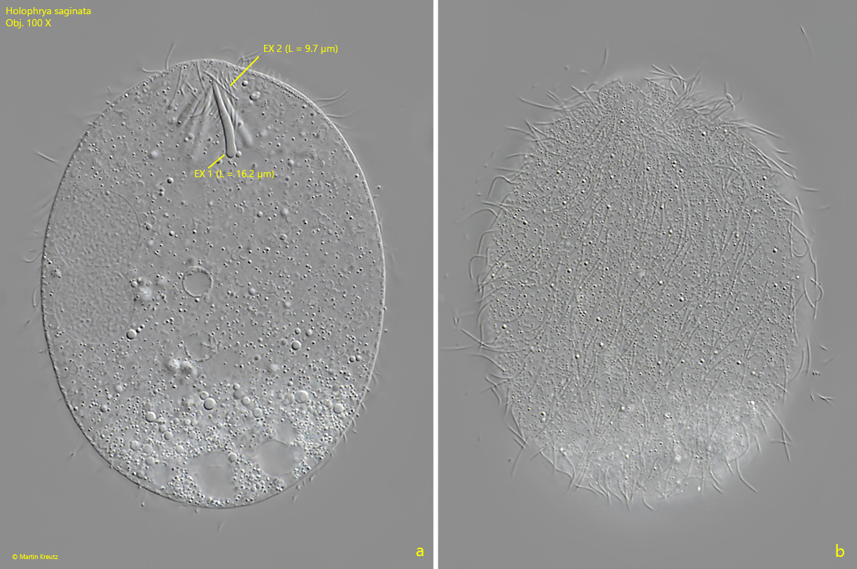
Holophrya saginata can be easily identified by the very large extrusomes surrounding the apical pharyngeal basket. The tapered tip of these extrusomes always points anteriorly. In some cases, these large, comma- and spindle-shaped extrusomes are also found scattered throughout the cytoplasm. These large extrusomes were 15–17 µm long in my specimens (s. figs. 3 a and 4). The pharynx is lined with a second variety of extrusomes, which are thin and rod-shaped (s. figs. 2 a-b and 3 a). According to my measurements this type of extrusomes is 9–10 µm long (s. fig. 3 a).
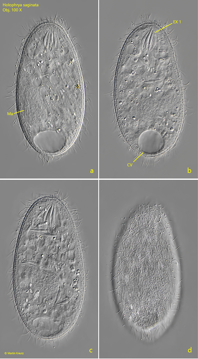
Fig. 1 a-d:Holophrya saginata. L = 86 µm. A freely swimming specimen. Note the large extrusomes (EX 1) surrounding the pharyngeal basket. CV = contractile vacuole, Ma = macronucleus. Obj. 100 X.
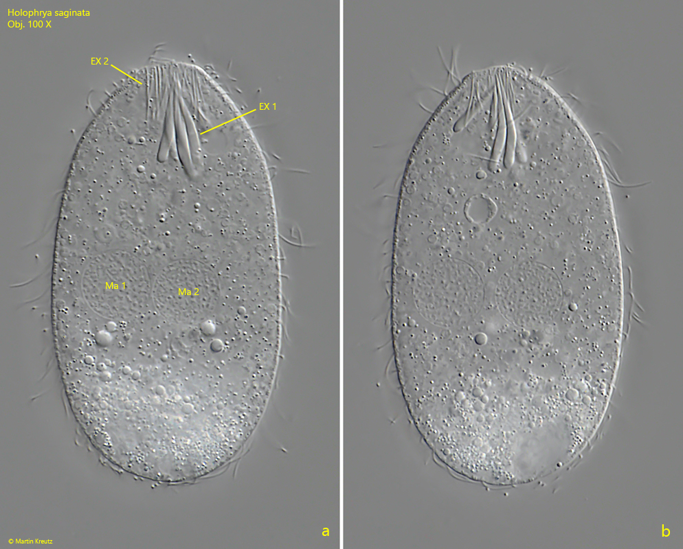
Fig. 2 a-b:Holophrya saginata. L = 74 µm. A slightly squashed second specimen. The pharyngeal basket is lined with thin rod-shaped extrusomes (EX 2) and surrounded by large, comma- and spindle shaped extrusomes (EX 1). This specimen has two macronuclei (Ma 1, Ma 2), probably due to a conjugation before. Obj. 100 X.
Fig. 3 a-b:Holophrya saginata. L = 74 µm. The squashed specimen as shown in fig. 2 a-b. The thin, rod-shaped extrusomes (EX 2) are 9–10 µm long and the comma-shaped extrusomes (EX 1) are 15–17 µm long. Obj. 100 X.
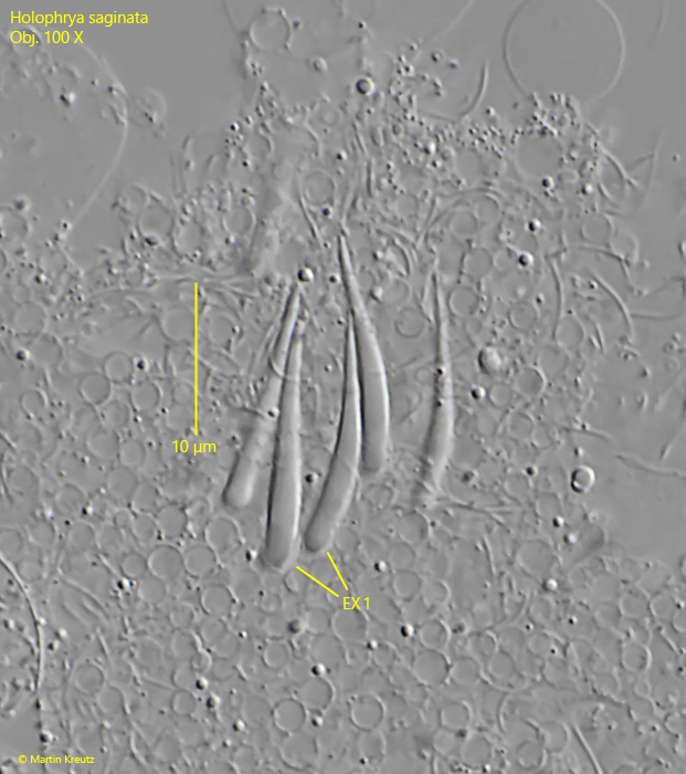
Fig. 4:Holophrya saginata. The comma-shaped extrusomes (EX 1) in detail. In this specimen these extrusomes are 15.8–16.3 µm long. Obj. 100 X.