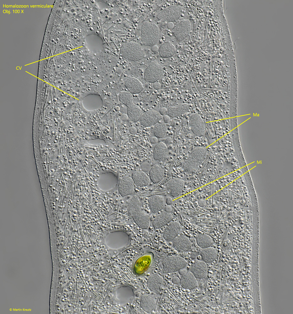
macronucleus moniliform of 20–50 nodes, about 25–50 spherical micronuclei
5–21 contractile vacuoles arranged in a row along dorsal side
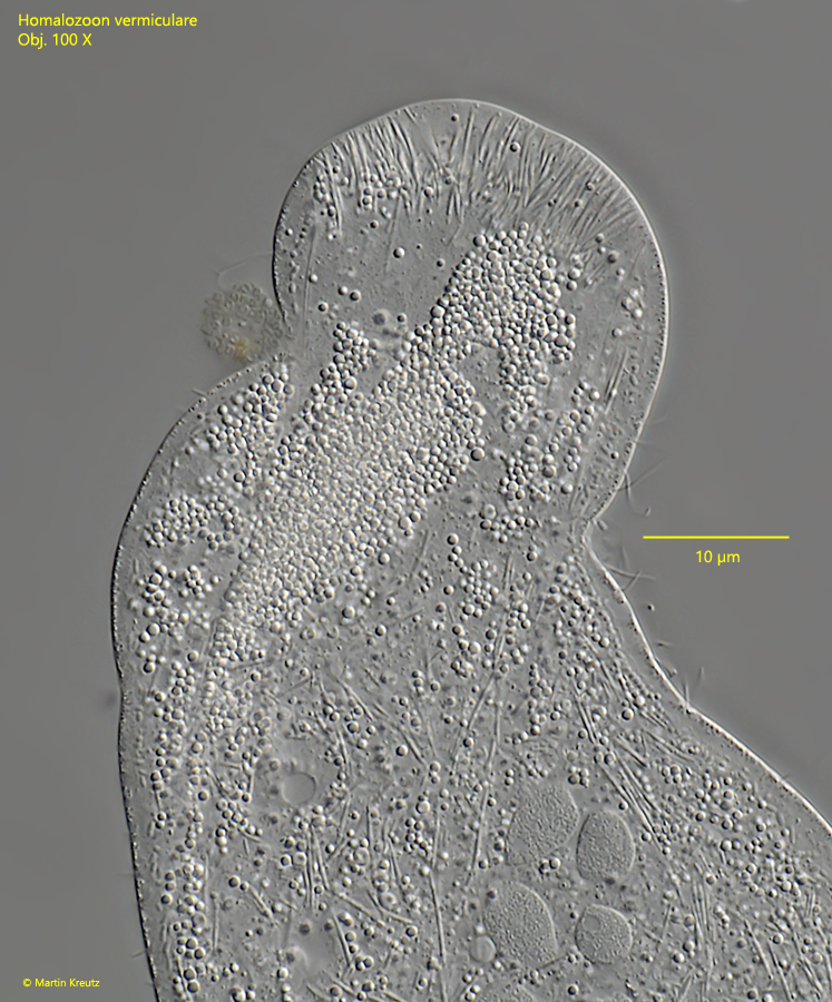
oral bulge almost hemi-spherically shaped with 2 µm long and 5–15 µm long extrusomes (rod-shaped)
below oral bulge a so-called parapharyngeal mass of small granules, sometimes colored yellowish
right side ciliated with 10–20 rows of cilia
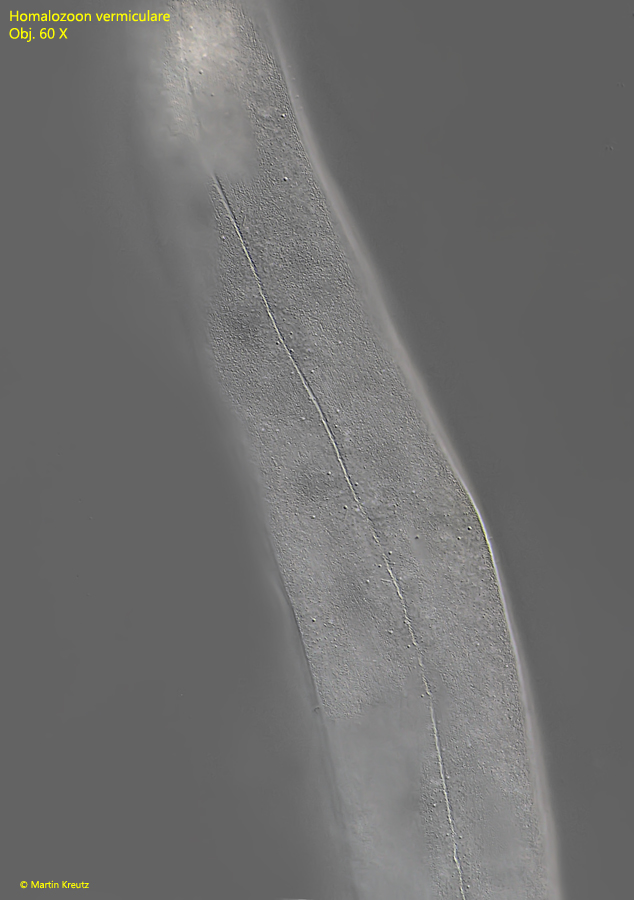
left side almost naked apart from 3–4 rows of short bristles and the dorsal brush
movement creeping, meandering, burrowing in detritus, rarely swimming
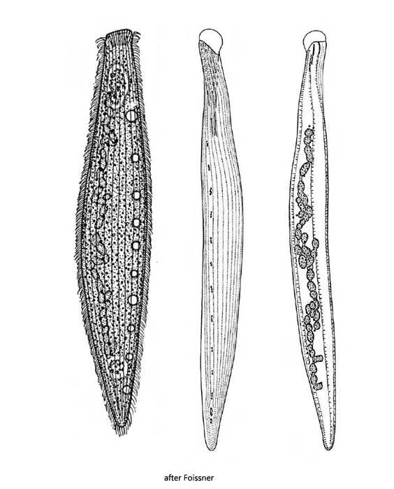
Homalozoon vermiculare
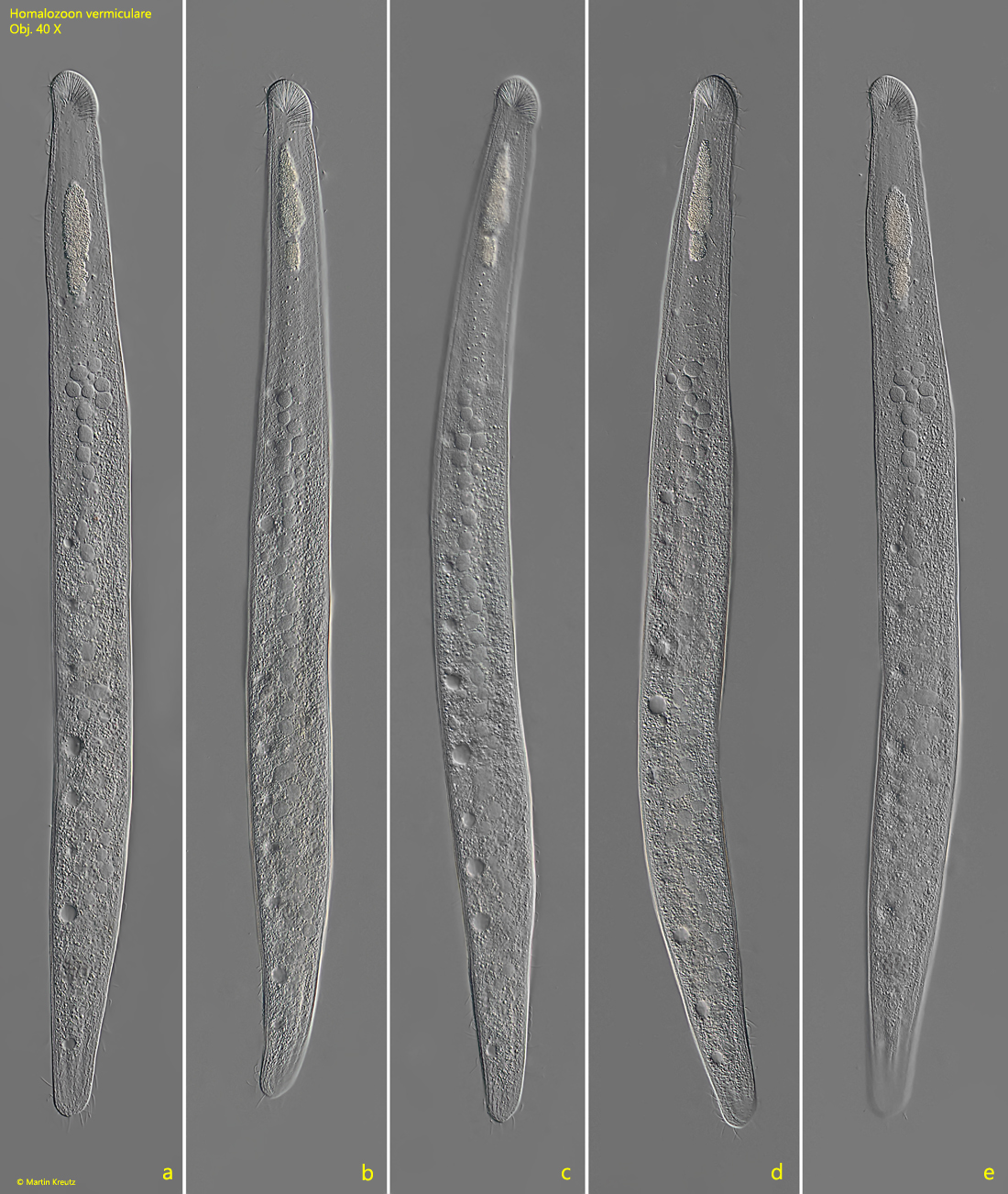
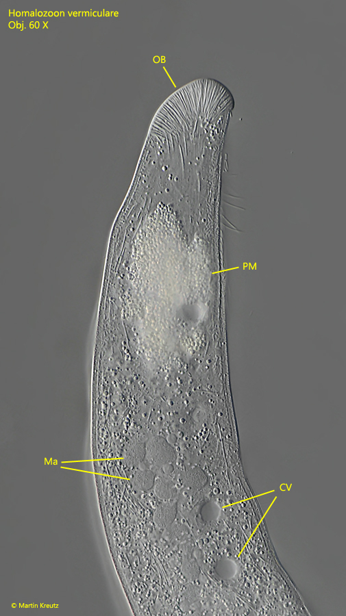
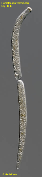
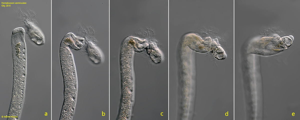
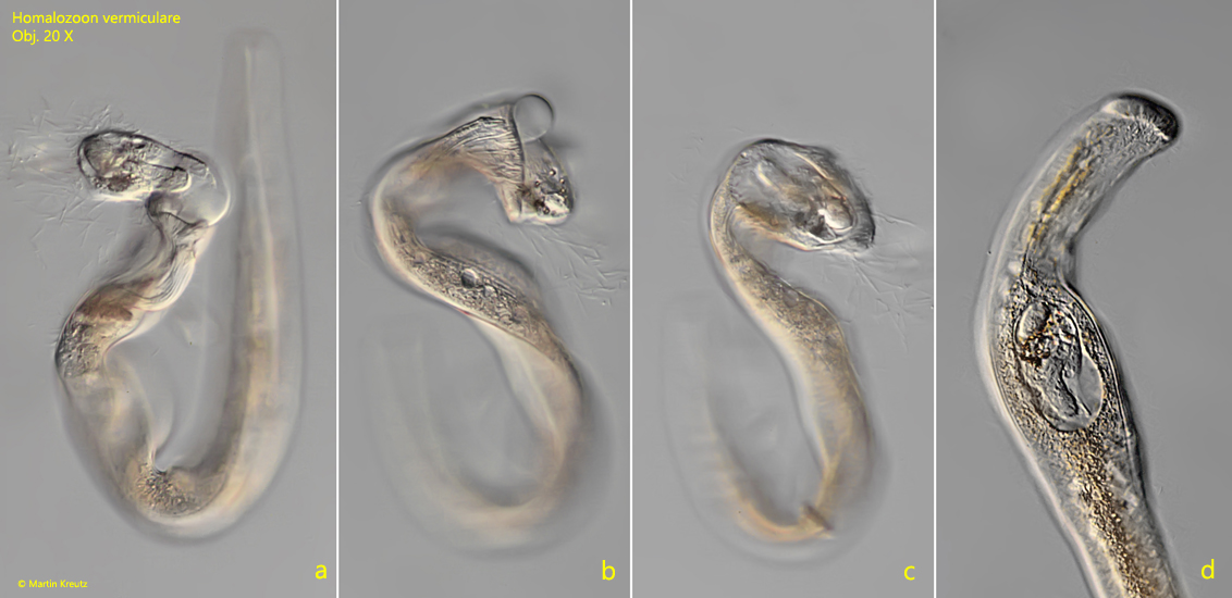
Homalozoon vermiculare is a large, characteristic ciliate that can hardly be confused with any other species. The body is slender and worm-like (s. fig. 1 a-e). In my population the specimens were about 500 µm long. The oral bulge is dome-shaped and densely covered with extrusomes (s. fig. 3). The moniliform macronucleus consists of 20–50 nodules, and is accompanied by numerous micronuclei (s. fig. 5). In parallel to the macronucleus is a row of 5–21 contractile vacuoles (s. fig. 5). The posterior end is tapered and rounded. A distinctive feature of Homalozoon vermiculare is a so-called parapharyngeal mass, which is located below the mouth opening (s. figs. 2, 3 and 4). It is often yellowish and has a granular character. It is described to consist of paraglycogen and various minerals. The exact purpose of the paraphyrangeal mass is not known. It is thought to play a role in the digestion of prey organisms, as it enters the food vacuole along with the prey when swallowed.
I was able to observe Homalozoon vermiculare best on floating coverslips, where the ciliate likes to settle when a fauna of prey organisms (ciliates, rotifers) has already formed there. The specimens then glide along the coverslip with the right, ciliated side.
Fig. 1 a-e: Homalozoon vermiculare. L = 495 µm. A freely gliding specimen. Obj. 40 X.
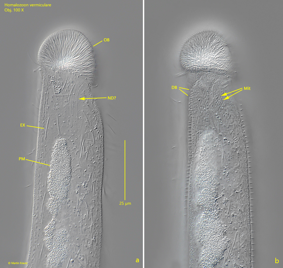
Fig. 2 a-b: Homalozoon vermiculare. Two focal planes of the oral bulge (OB) of a freely swimming specimen. DB = part of the dorsal brush, EX = extrusomes, Mit = mitochondria, ND? = probably nemadesmata, PM = parapharyngeal mass. Obj. 100 X.
Fig. 3: Homalozoon vermiculare. A slightly squashed specimen. Note the parapharyngeal mass (PM) below the oral bulge (OB). CV = contractile vacuoles. Ma = nodes of the moniliform macronucleus. Obj. 60 X.
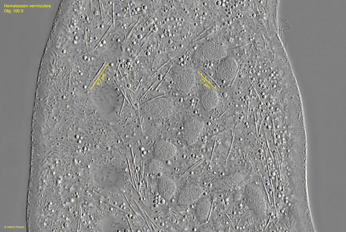
Fig. 3: Homalozoon vermiculare. The parapharyngeal mass is an accumulation of small spherules with a diameter of about 1 µm. Obj. 100 X.
Fig. 5: Homalozoon vermiculare. The nodules of the moniliform macronucleus (Ma) with the adjacent micronuclei (Mi) in a strongly squashed specimen. Obj. 100 X.
Fig. 6: Homalozoon vermiculare. In the cytoplasm slightly curved, rod-shaped extrusomes with a length of about 8 µm are scattered. Obj. 100 X.
Fig. 7: Homalozoon vermiculare. The left side is almost naked apart from 3-4 rows of short bristles. The bristles are located in furrows. Obj. 100 X.
Fig. 8: Homalozoon vermiculare. A specimen in cell division. Obj. 10 X.
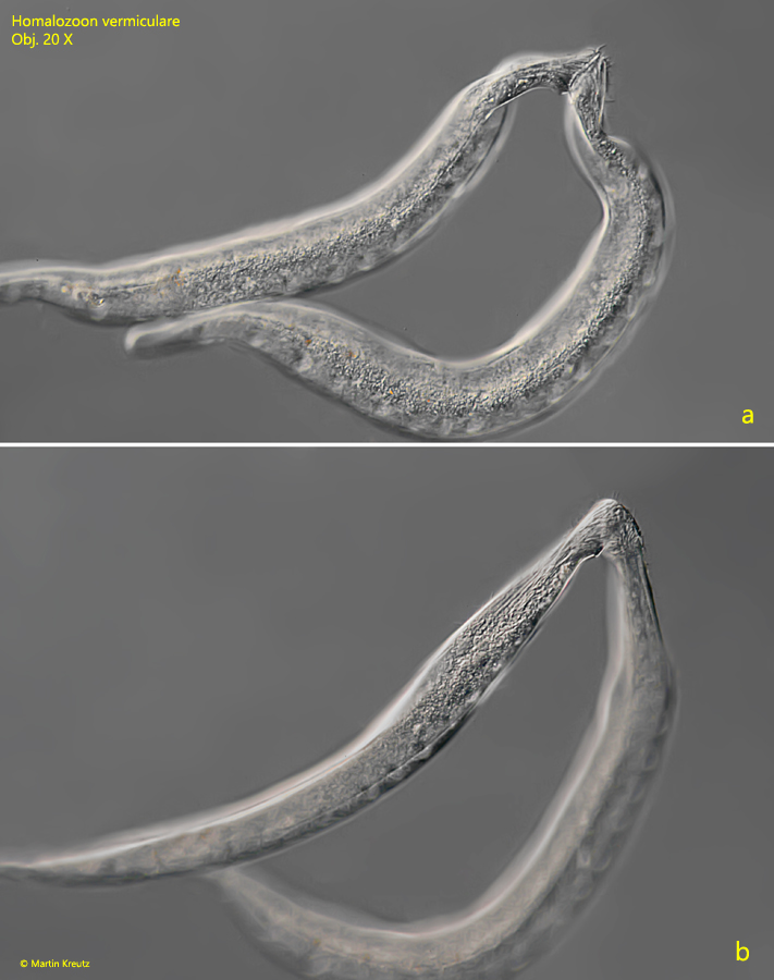
Fig. 9 a-b: Homalozoon vermiculare. Two specimens in conjugation connected via the mouth openings. Obj. 20 X.
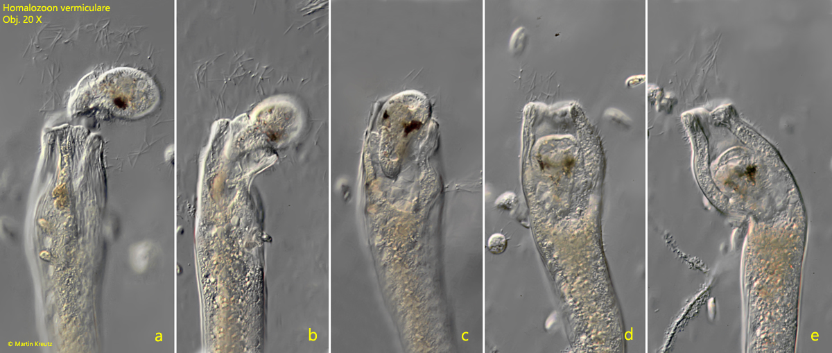
Homalozoon vermiculare is a voracious predator that especially likes to feed on ciliates. It also phagocytoses species that have extrusomes, such as Frontonia or Paramecium. I was able to observe prey capture several times in microaquaria (s. figs. 10 a-e, 11 a-d and 12 a-e). After contact of the prey with the oral bulge of Homalozoon vermiculare, the prey ejects a cloud of extrusomes as a reaction. I could not see the extrusomes ejected by Homalozoon vermiculare, however, the prey ciliate immediately begins to denature and is unable to move. Homalozoon vermiculare then immediately begins phagocytosis. The process takes about 30 seconds to 1 minute before the prey is completely swallowed.
Fig. 10 a-e: Homalozoon vermiculare. A specimen attacks a ciliate and devours it. Note the cloud of extrusomes ejected by the prey. Obj. 20 X.
Fig. 11 a-d: Homalozoon vermiculare. A second specimen attacks a ciliate and devours it. Obj. 20 X.
Fig. 12 a-e: Homalozoon vermiculare. A third specimen attacks a ciliate and devours it. Obj. 20 X.