one oval macronucleus with one globular micronucleus attached to macronucleus
cortex with mucilaginious layer with lepidosomes
a conspicuous flagellum-like, retractable mouth process
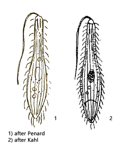
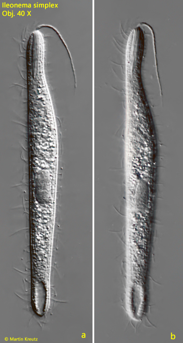
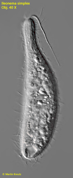
Ileonema simplex
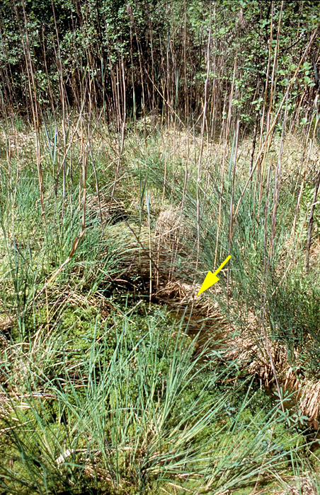
The first describer Eugène Penard found only one specimen of Ileonema simplex in 1916, on which his drawing and description is based. Kahl later did not find this species himself, but took over the drawing and description from Penard. In November 1999 I discoverd Ileonema simplex in a small brown water puddle located in Simmelried, which was overgrown with Sphagnum moss (assigned as pond 4, s. Simmelried).
Fig. 1: Ileonema simplex. The brown water puddle in Simmelried where Ileonema simplex was found between 1999–2000.
Most of the specimens were found in squeezed Sphagnum moss. After a few days at room temperature, small flakes, about 2 mm in size, floated near the water surface of the samples, harboring an extraordinary diversity of species from ciliates, flagellates and rotifers. Among them, Ileonema simplex was found in large numbers. About 50 specimens were present in one of these flakes. After the year 2000 the species disappeared again and no more specimens could be found since then.
Due to the flagellum-like mouth process Ileonema simplex cannot be misidentified. This mouth flagellum is clearly visible even at lower magnifications. It has an average length of 30–40 µm. It tapers evenly towards the end. However, under no circumstances the mouth flagellum is twisted as Kahl describes it for Ileonema dispar. A confusion with Ileonema ciliata can also be excluded, because this species has two macronuclei.
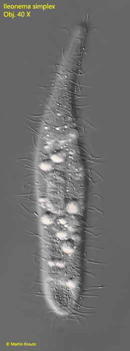
Fig. 3: Ileonema simplex. L = 140 µm (without mouth flagellum), a more contracted, freely swimming specimen. Obj. 40 X.
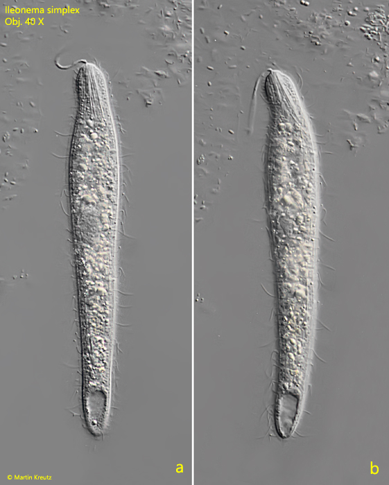
Fig. 4 a-b: Ileonema simplex. L = 181 µm (without mouth flagellum). This specimen is burrowing in a detritus flake. Obj. 40 X.
Since many specimens of Ileonema simplex were available in the locality, a morphometric survey could be performed. The survey was done with the 40 X objective and exclusively on freely swimming specimens. Only the measurement of the macronucleus had to be done on specimens with little squeezing. The results are summarized in table 1.
Tab 1: Morphometric data of Ileonema simplex.
Parameter
n
Average
(µm)
Min
(µm)
Max
(µm)
Body length
28
141
98
191
Body width
28
26
15
34
Macronucleus
length
18
18
15
22
Macronucleus
width
18
11
20
22
Length
mouth flagellum
10
37
20
54
I was able to examine the mouth flagellum in detail. It appears to be made of excreted mucus or plasm. It is by no means actively motile and may also be retracted, which can be observed with increasing pressure on the coverslip. At the origin, the organelle is smooth and homogeneous, but may be dissected toward the distal end. Moreover, it appears that these segmented mucus secretions are attached to a kind of filament. Despite intensive observation of about 100 specimens, no clues emerged as to the function of the organelle, such as prey capture. A tactile function seems to be out of question. I could often observe touching of the mouth flagellum by potential prey organisms (ciliates, flagellates), but this remained without any reaction. Ileonema simplex swims slowly and is very flexible. The ciliate penetrates cracks and cavities of detritus flakes and Zoogloea flakes, in which it moves back and forth. In any case, during this searching movement the flagellum is only passively dragged along and there is nothing to indicate a sensory function. Also what Ileonema simplex feeds on could not be determined.
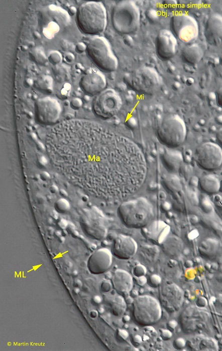
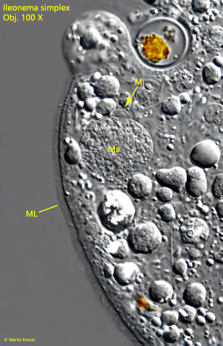
Ileonema simplex is member of the family Trachelophyllidae. Ciliates in this family are covered with a mucilaginious layer containing lepidosomes (epicortical scales). The lepidosomes of Ileonema simplex cannot be seen in detail with light microscopy but at higher magnification the mucilaginuous layer appears “rough” by the lepidosomes.
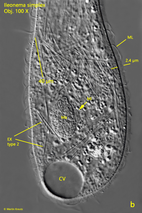
Fig. 5 + 6: Ileonema simplex. The mucilaginuous layer (ML) is about 2.5 µm thick. Ma = macronucleus; Mi = micronucleus. Obj. 100 X.
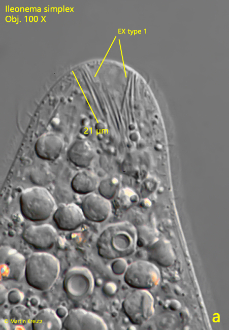
The oral bulge of Ileonema simplex is equipped with a bundle of 21 – 24 µm long, rod-shaped extrusomes. In the plasm of some specimens a second type of rod-shaped extrusomes with a length of about 40 µm was detected.
Fig. 7 a-b: Ileonema simplex. In the oral bulge a bundle of rod-shaped extrusomes with a length of 21 – 24 µm is present (a) and in the plasm rod-shaped extrusomes with a length of about 40 µm (b). EX = extrusomes, Ma = macronucleus, Mi = micronucleus, ML = mucilaginuous layer. Obj. 100 X.
Penard mentions in his original description of Ileonema simplex that the widely spaced cilia would be longer at the anterior and posterior ends of the ciliate than in the mid-body. I cannot confirm this. The cilia are of the same length over the whole body (about 15 µm).
Fig. 8: Ileonema simplex. The widely spaced cilia are soft with a constant length of 15–18 µm over the whole body. Obj. 40 X.