body shape variable, hyaline dactylopodia finger-shaped or long conical
uroid absent
length 70–100 µm
one spherical nucleus with central nucleolus
one contractile vacuole
no crystals in cytoplasm
cell covered with a coat of scales (visible in electron microscope)
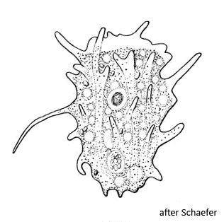
Korotnevella bulla
Korotnevella bulla was originally described as Mayorella bulla by Schaeffer in 1926. In 1982, Page transferred Mayorella bulla to the genus Dactylamoeba. At the suggestion of Goodkov, the genus Dactylamoeba was renamed Korotnevella in 1988 (Korotneff created the genus Dactylamoeba in 1880). Therefore, the current name of this amoeba is Korotnevella bulla.
I find Korotnevella bulla frequently and regularly in the Simmelried. All specimens were larger than 80 µm and thus larger than the similar Korotnevella stella (31-60 µm). Therefore, I do not believe that this is a large form of Korotnevella stella.
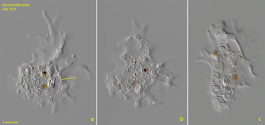
Korotnevella bulla flows forward with finger-shaped psudopodia ( = dactylopodia). The cytoplasm is very transparent and contains no inclusions apart from some food vacuoles. A contractile vacuole is present and the broadly oval nucleus with a central nucleolus (s. fig. 3). An uroid is not formed during locomotion.
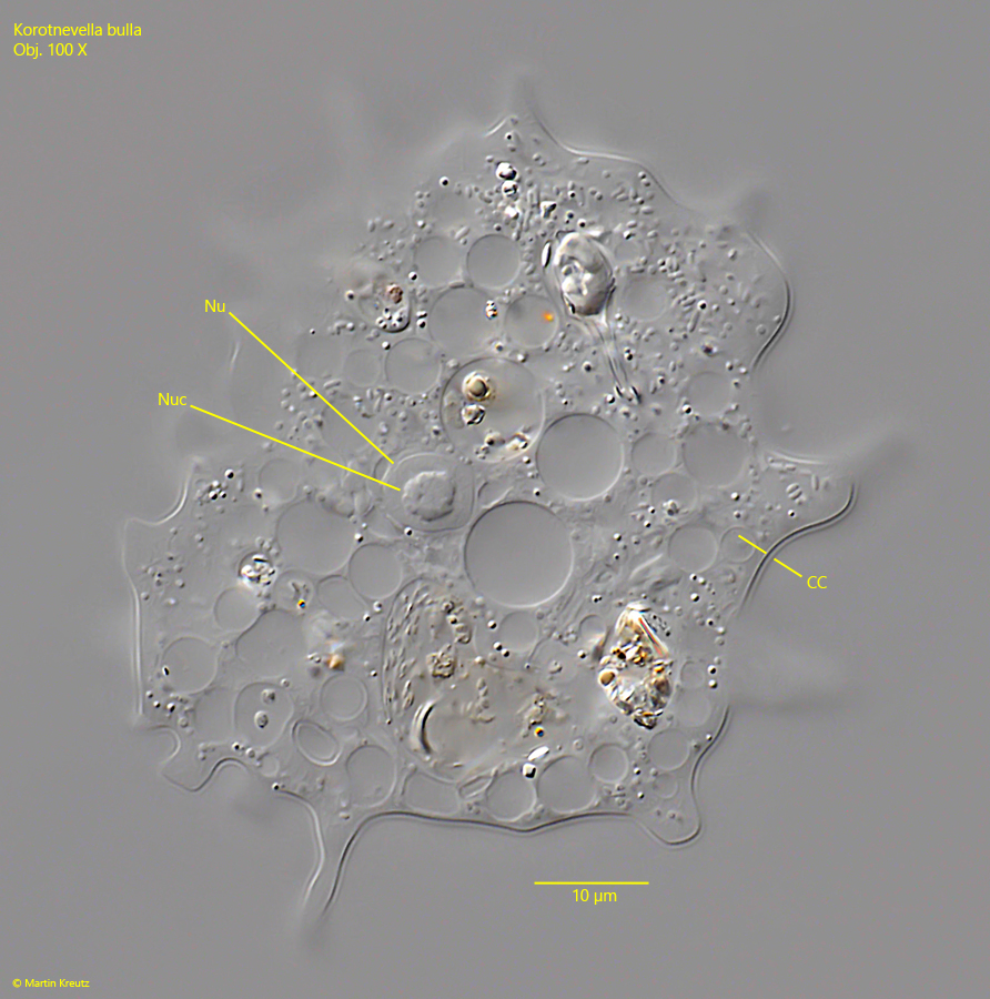
The species of the genus Korotnevella are covered with a layer of scales, the so-called cell coat. The shape of the scales can only be seen under an electron microscope and were studied thouroughly by Voelker & Clauss. Under the light microscope, however, this cell coat can be seen as a thin film and, under favorable conditions, a structuring of this layer at the limit of the possible resolution can also be seen (s. fig. 4).
Fig. 1 a-c:Korotnevella bulla. L = 102 µm. A freely floating specimen. CV = contractile vacuole. Obj. 40 X.
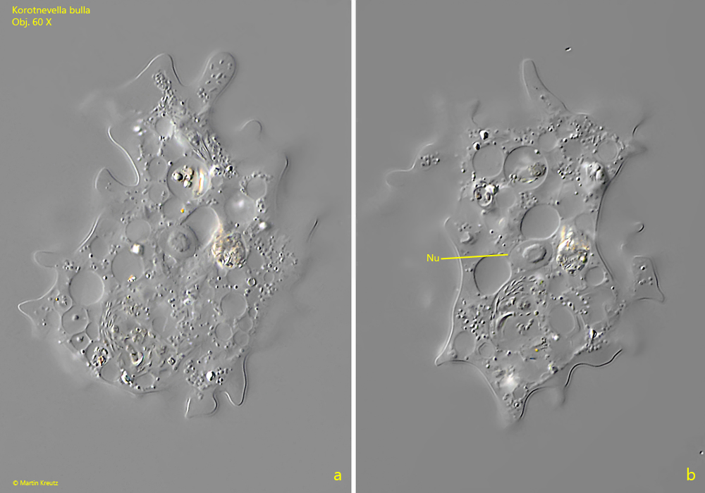
Fig. 2 a-b:Korotnevella bulla. L = 84 µm. A second specimen. Nu = nucleus. Obj. 60 X.
Fig. 3:Korotnevella bulla. The squashed specimen as shown in fig. 2 a-b. Note the thin cell coat (CC) covering the specimen. This coat contains < 0.1 µm scales, only visible in the electron microscope. Nu = nucleus, Nuc = central nucleoli. Obj. 100 X.
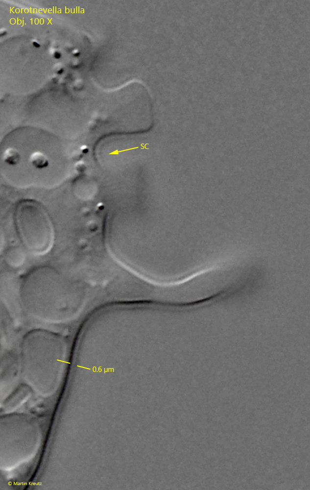
Fig. 4:Korotnevella bulla. A strongly enlarged and contrasted section of the cell coat. At the limit of possible resolution, a structure of the cell coat can be recognized, which is caused by the scales (SC) embedded in it. The cell coat is 0.5–0.6 µm thick. Obj. 100 X.