marginal cells occasionally bearing tufts of fine fibers at tips of lobes
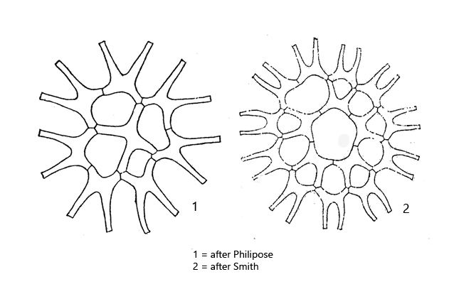
the intercellular gaps are large and occupy as much space as the cells
one parietal chloroplast
single pyrenoid
Lacunastrum gracillimum
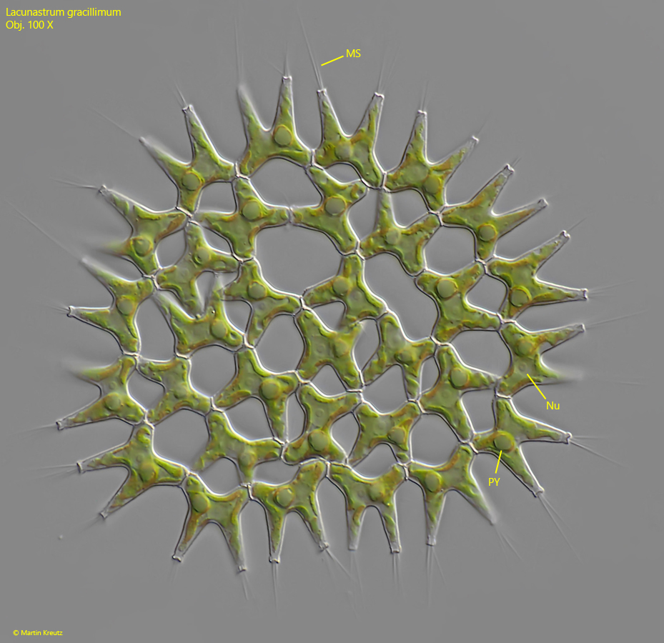
I found Lacunastrum gracillimum in the plankton of the strongly eutrophic pond of the waste disposal company Constance. This pond is fed by the purified water of the sewage plant, which is still very rich in nutrients. Lacunastrum gracillimum can be easily recognized by the delicate, H-shaped cells and the large gaps between the cells, which occupy as much space as the cells. The cells on the outer margin each bear two long projections. At the distal ends of the projections tufts of thin fibers are visible, which I interpret as an adaptation to the planktonic habitat.
The species Pediastrum duplex var. gracillimum was transferred to Lacunastrum gracillimum by McManus in 2011.
Fig. 1: Lacunastrum gracillimum. D = 92 µm. A slightly squashed specimen. MS = mucilaginous spines, Nu = nucleus, PY = pyrenoid. Obj. 100 X.
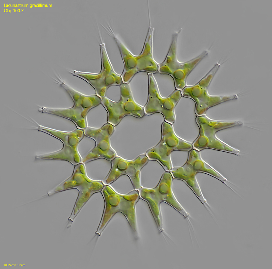
Fig. 2: Lacunastrum gracillimum. D = 65 µm. A slightly squashed second specimen. Obj. 100 X.