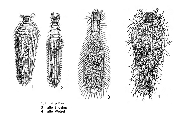
body flask-shaped, at anterior end (“neck”) with 3–5 annular furrows
anterior neck can be retracted
cell surface with longitudinal furrows
length 130–200 µm, width 40–60 µm
macronucleus ellipsoid or bean-shaped, one ellipsoid micronucleus
contractile vacuole at posterior end
37–50 longitudinal rows of cilia
in the cytoplasm rod-shaped, thin extrusomes, about 15 µm long
oral bulge with about 30 chevron-shaped rods
Lagynus elegans
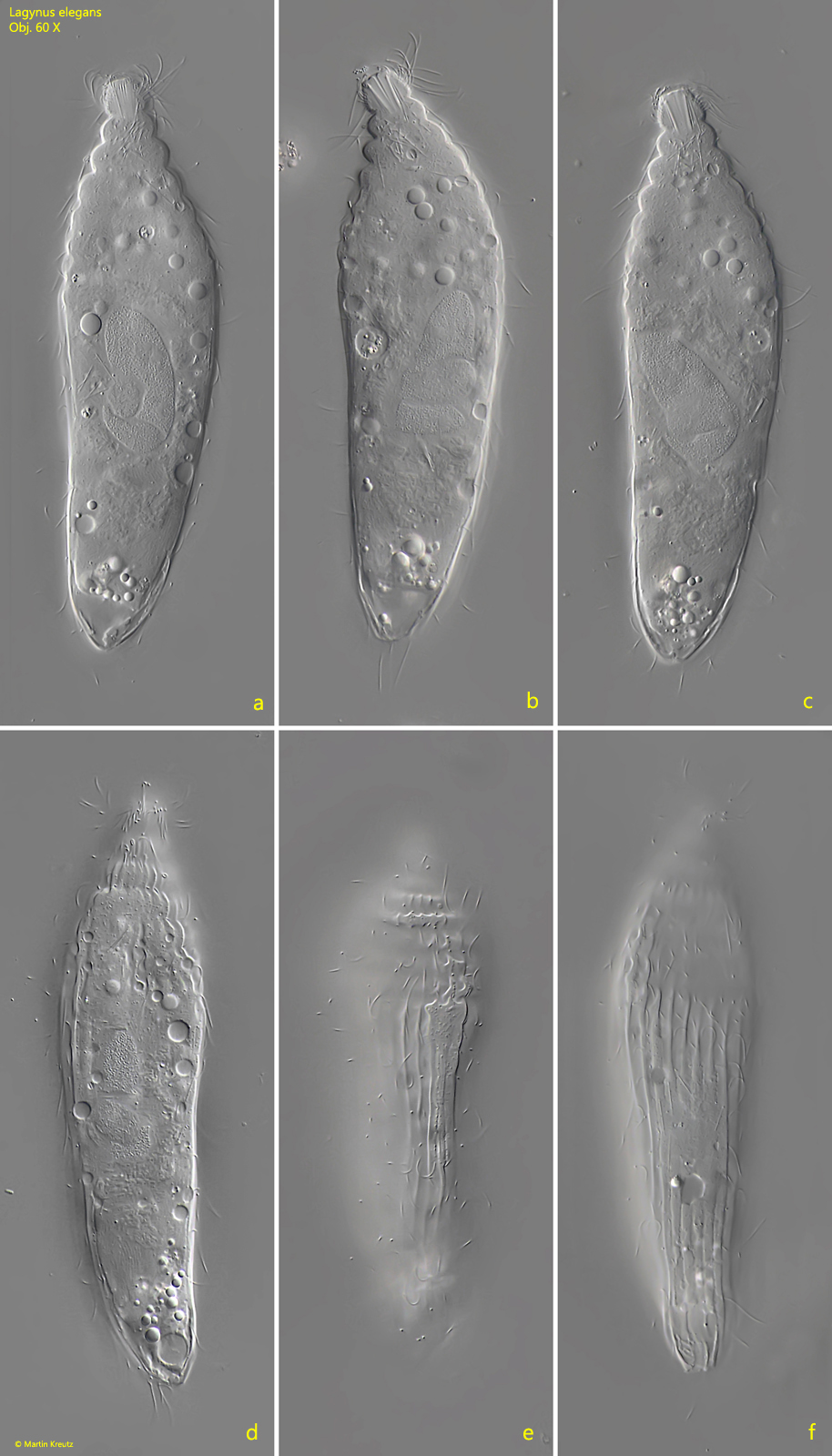
I find Lagynus elegans rarely, but regularly. In some years the species appeared with 5–10 specimens per milliliter. The species is easy to recognize by the stepped neck, which is caused by ring-shaped furrows. There should be 3–5 furrows. However, at high magnification I had the impression that there are 7 furrows, although the furrows become less pronounced towards the posterior end (s. fig. 7). The longitudinal furrows of the pellicle (s. figs. 1 e, 1 f and 7) are not mentioned by either Kahl or Foissner, although I was able to detect them in all specimens in my population. The extrusomes scattered in the cytoplasm should be about 15 µm long according to Foissner. In my specimens I could measure lengths between 9–11 µm.
According to Foissner the species Lagynus elegans should be synonymous with Lacrymaria sapropelica. After I having found a Lacrymaria which has the characteristics of Lacrymaria sapropelicadescribed by Kahl (e.g. head broader than the neck), I am convinced that Lacrymaria sapropelica is a distinct species which is not synonymous with Lagynus elegans. Since I was able to detect both species in my sites, I was able to make a direct comparison. Lacrymaria sapropelica not only has a differently shaped head and neck, but is also smaller than Lagynus elegans with about 90 µm, does not have the conspicuous longitudinal furrows on the pellicle and has fewer ciliary rows.
Fig. 1 a-f:Lagynus elegans. L = 142 µm. Different focal planes of a freely swimming specimen. Obj. 60 X.
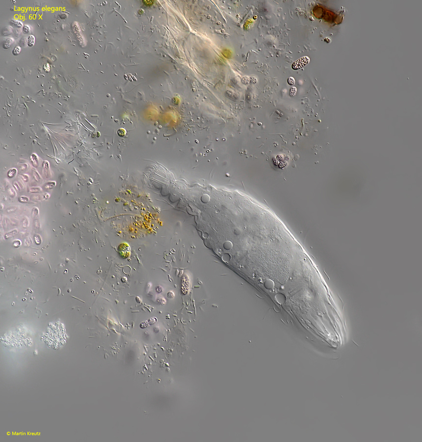
Fig. 2:Lagynus elegans. L = 142 µm. The specimen shown in fig. 1 a-f burrowing in a detritus flake. Obj. 60 X.
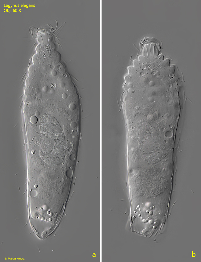
Fig. 3 a-b:Lagynus elegans. L = 135 µm. A specimen with elongated (a) and retracted neck (b). Obj. 60 X.
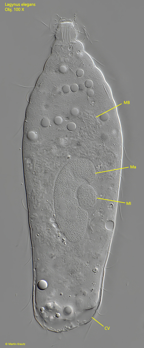
Fig. 4:Lagynus elegans. L = 148 µm. Total view of a squashed specimen. CV = contractile vacuole, Ma = macronucleus, MB = intracellular mass of bacteria?, Mi = micronucleus. Obj. 100 X.
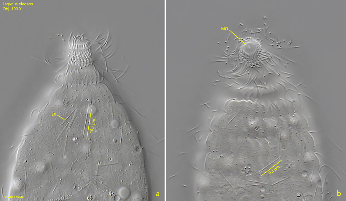
Fig. 5 a-b:Lagynus elegans. Lateral (a) and apical view (b) of the head. Note the thin, rod-shaped extrusomes (EX) scattered in the cytoplasm. MO = mouth opening. Obj. 100 X.
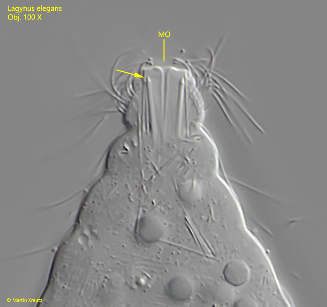
Fig. 6:Lagynus elegans. The oral bulge in detail. It seems that the pharyngeal extrusomes (arrow) have tapered ends with a tip. MO = mouth opening. Obj. 100 X.
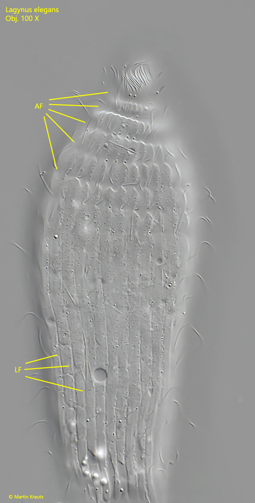
Fig. 7:Lagynus elegans. Anteriorly the end tapers to the distal end by annular furrows (AF). Caudal to these furrows, longitudinal furrows (LF) run in parallel to the posterior end. Obj. 100 X.
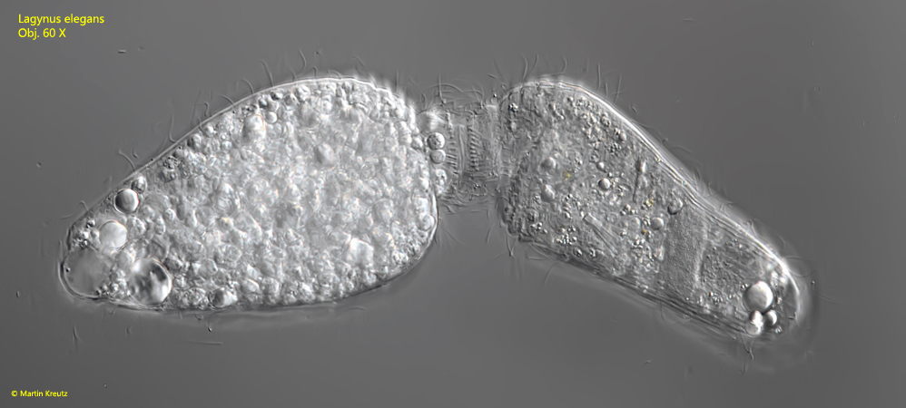
Fig. 8:Lagynus elegans. L = 128 µm + 118 µm. Two specimens in conjugation. Obj. 60 X.