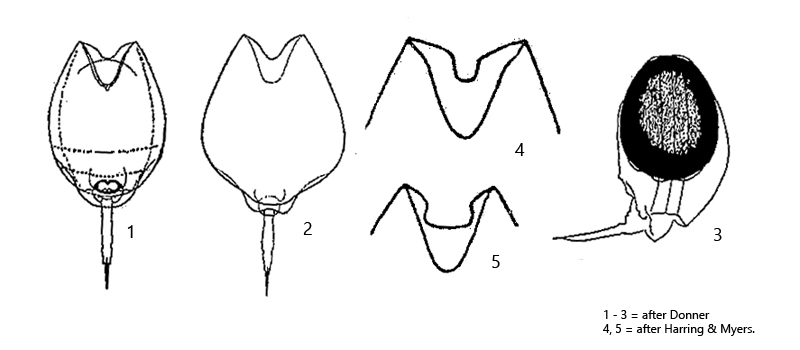
anterior ventral margin of lorica with a deep V-shaped incision
anterior dorsal margin of lorica with a more shallow incision
length 170 -180 µm
posterior body segment shield-shaped, rounded and covering foot
toe very long, claws divided with distinct basal spicules
one eyespot
resting egg with smooth surface, colored brown
Lecane bulla
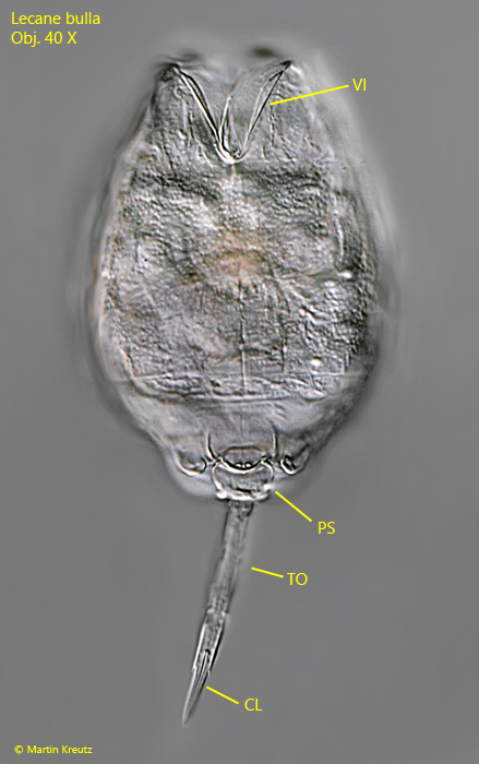
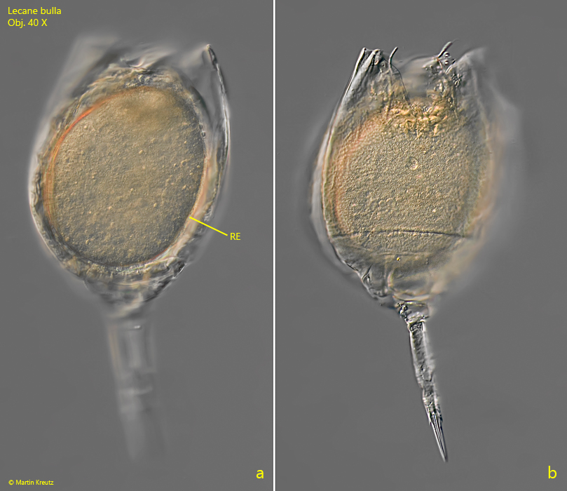
I have found Lecane bulla so far exclusively in the Simmelried between floating plants. The findings were made in October 2000 and November 2007. Lecane bulla can be recognized quite easily by the deep, V-shaped incision at the anterior ventral margin of the lorica. At the posterior end of the ventral side a round shield-shaped plate is visible (s. fig. 1), which covers the foot (which is thus not visible). The toe is long and ends in two pointed claws. During the reproductive cycle, one resting egg is formed per individual before winter. The large resting egg is not laid, but remains in the lorica after the mother dies (s. drawing 3 above and fig. 2 a-b). The resting egg is released only after decomposition of the mother’s lorica.
Fig. 1:Lecane bulla. L = 200 µm (with toe amd claws). Ventral view of a slightly squashed specimen. Note the V-shaped incision of the anterior ventral margin of the lorica (VI). CL = claws, PS = posterior shield-shaped segment, TO = toe. Obj. 40 X.
Fig. 2 a-b:Lecane bulla. L = 200 µm (with toe amd claws). Two focal planes of a dead specimen with a resting egg (RE) remaining in the lorica. Obj. 40 X.