cell oval or bag-shaped, anterior end laterally flattened
CV terminal
macronucleus vermiform
One globular micronucleus attached to the macronucleus
each 16 tentacles on dorsal and ventral side of body
tentacles restricted to posterior third of body
tentacles armed with extrusomes and with a distal ring of cilia
dorsal brush up to a quarter of the body length
Legendrea loyezae
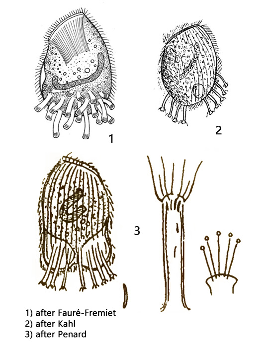
The first description of Legendrea loyezae was made by Fauré-Friemet in 1908. After this the species was also found by Penard (1914) and last by Kahl (1935), but always only sporadically. There are no records after 1935. I found Legendrea loyezae in May 2014 first in the Ulmisried and later several specimens in the Simmelried and the Purren pond (Aug 2017 and Oct 2021). However, I always found only isolated specimens in the upper layer auf decayed leaves. The appearance of Legendrea loyezae is unmistakable because the ciliate has bundles of tentacles arising from its posterior end. The swimming style is sluggish and slowly rotating.
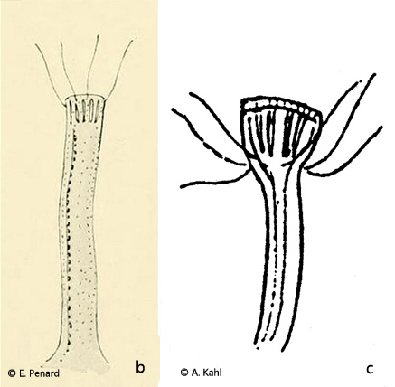
The ciliate is ovoid in shape, with an oblique, about 40 µm long and 5 µm wide oral bulge. The anterior end is laterally flattened. While Fauré-Fremiet and Penard still assumed a lateral arrangement of the tentacles, Kahl describes the origin of the tentacles as ventral and dorsal from a notch-like depression, which also encircles the posterior end of the ciliate. I could confirm this arrangement described by Kahl. Therefore, from a ventral or dorsal view, Legendrea loyezae appears to terminate at the posterior end in two saccular projections. The contractile vacuole in my specimen was found in the right posterior convexity. The number of tentacles seems to be constant. I could count 32, each 16 on the dorsal and ventral side.
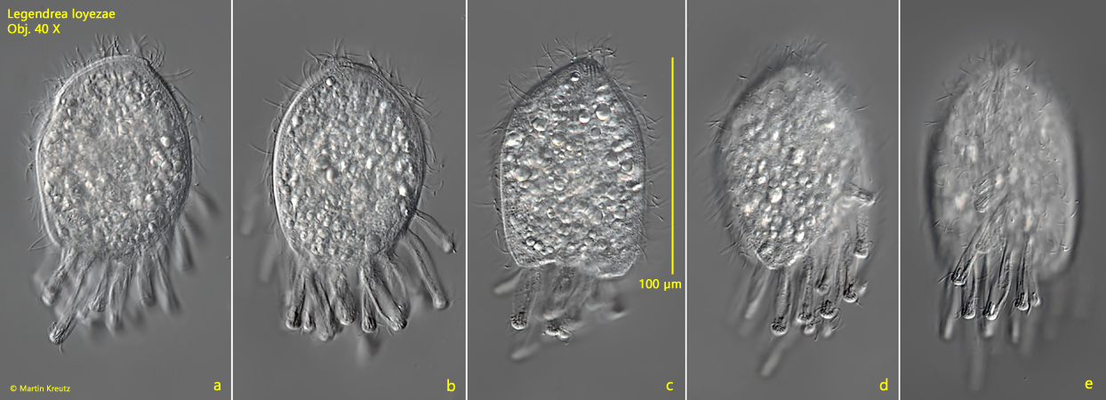
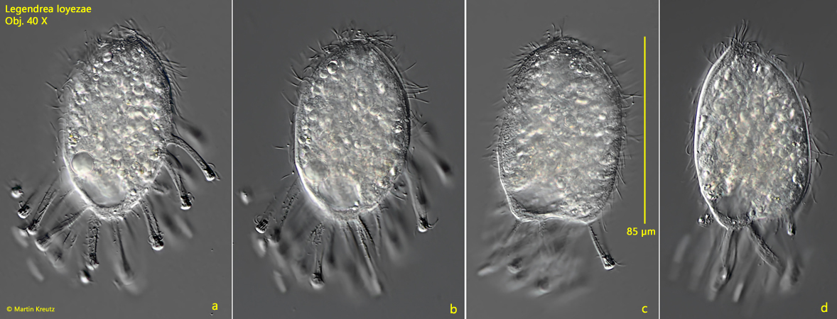
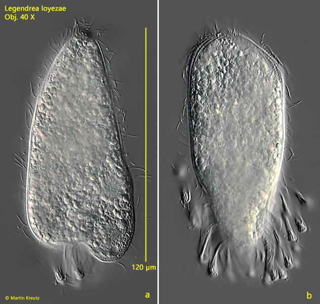
Fig. 2 a-d:Legendrea loyezae. L = 85 µm (without tentacles), a second freely swimming specimen. Obj. 40 X.
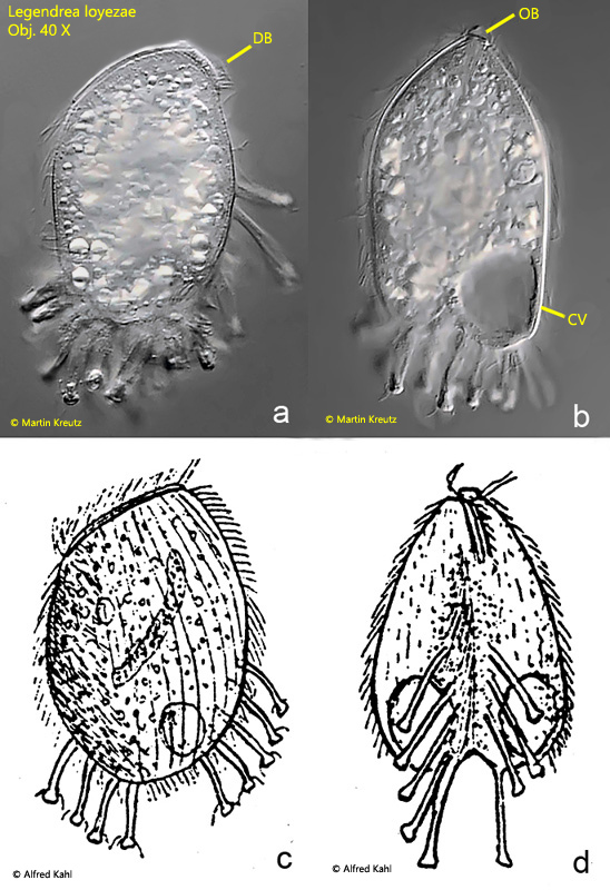
Fig. 3 a-d:Legendrea loyezae. Comparison of a freely swimming specimen with Kahl’s drawings. a, c) left side; b, d) dorsal view. DB = dorsal brush, OB = oral bulge. Obj. 40 X.
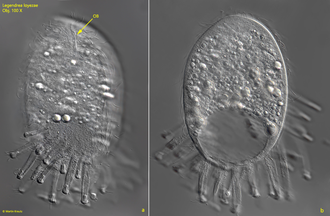
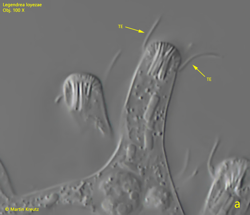
Fig. 4 a-b:Legendrea loyezae. A dorsal (a) and lateral (b) view of a slightly squashed specimen. OB = oral bulge; TE = tentacles. Obj. 100 X.
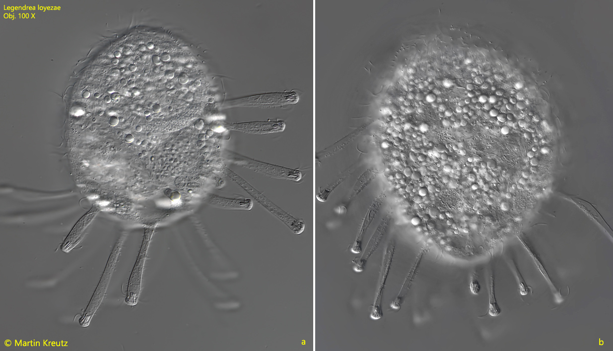
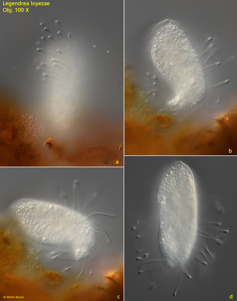
Fig. 5 a-b:Legendrea loyezae. A specimen with extended tentacles. 100 X.
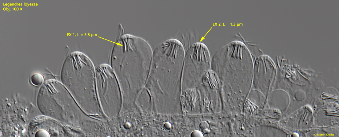
The structure of the tentacles of Legendrea loyezae has already been studied in great detail by Penard and Kahl. Distally, the tentacles end in papillae-shaped thickenings, in which 2 types of extrusomes are arranged. The distal, cap-shaped fringe is formed by very short extrusomes, only about 1 – 1.5 µm long, which are very densely arranged. Below them a bundle of about 10 slightly curved, 5 – 6 µm long extrusomes is arranged. The tentacles passively trailed behind when swimming. They were between 20 and 45 µm long and had a diameter of about 6 µm.
Fig. 6:Legendrea loyezae. Two types of extrusomes (Ex 1, Ex 2) in the tentacles of a squashed specimen. Obj. 100 X.
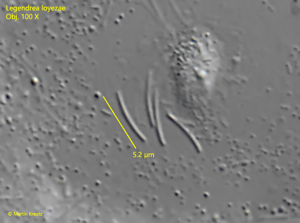
Fig. 7:Legendrea loyezae. The slightly curved extrusomes of the type 1 located in the distal ends of the tentacles. Obj. 100 X.
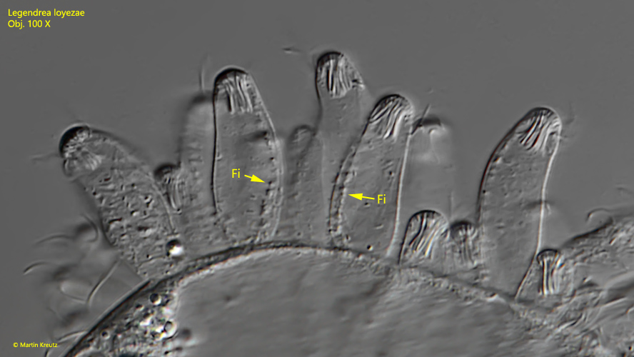
The tentacles themselves are hollow tubes. It appeared to me that they are protrusions of the pellicle. There appears to be some sort of a filament running in the tentacles between the base and the distal end. However, with higher magnification, one can see that this filament obviously rests on the inner tentacle wall and is made of fine granules, just as Penard drew it.
Fig. 8:Legendrea loyezae. The granular filaments on the inner side of the tentacles. Fi = filaments. Obj. 100 X.
The tentacles themselves are not ciliated, except for a circular arrangement of cilia just below the distal papilla. This arrangement differs somewhat from the observations of Penard and Kahl, who draw the origin of the cilia at the distal end (Penard) or below the bundle of the long extrusomes type 1 (Kahl).
Fig. 9 a-c:Legendrea loyezae. The annular cilia of the tentacles arises about 3 µm below the distal end (a) and not at the distal end (b, Penard) or below the bundle of extrusomes (c, Kahl). Obj. 100 X.
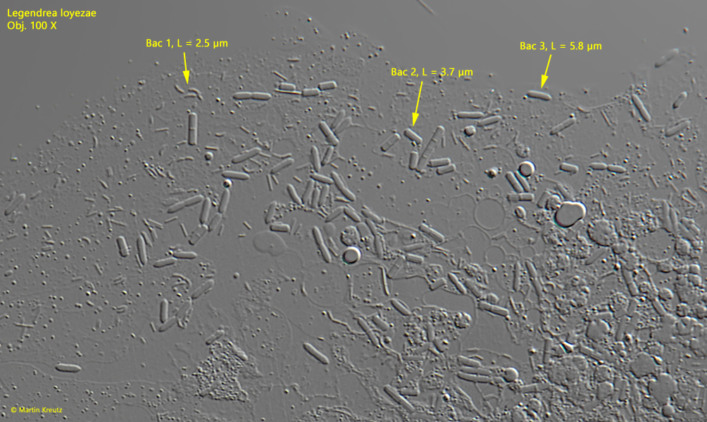
The plasma of Legendrea loyezae is filled with highly refractive, oil-like (reserve ?) bodies with a diameter of approx. 5 – 15 µm, which makes the ciliate completely opaque in the unsquashed state. In the squashed specimen, however, a great number of symbiotic bacteria were visible between these reserve bodies, composed of at least 3 species.
Fig. 10:Legendrea loyezae. There are at least 3 types of symbiotic bacteria present in the plasm. Bac 1 = curved rods, about 2.5 µm long, Bac 2 = cylindrically shaped rod, about 4 µm long, Bac 3 = rod with tapered ends, about 6 µm long. Obj. 100 X.
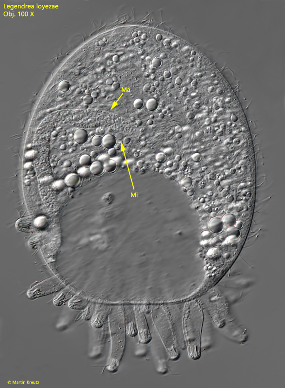
The macronucleus of Legendrea loyezae is vermiform and about 70 – 90 µm long. The globular micronucleus is hard to see. In a not completely squashed specimen it is found attached to the macronucleus.
Fig. 11:Legendrea loyezae. The globular micronucleus (Mi) is attached to the vermiform macronucleus (Ma). Obj. 100 X.
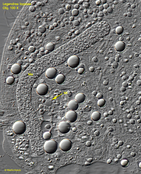
Fig. 12:Legendrea loyezae. In a more squashed specimen the micronuclus (Mi) is found to be detached from the macronucleus (Ma). Obj. 100 X.
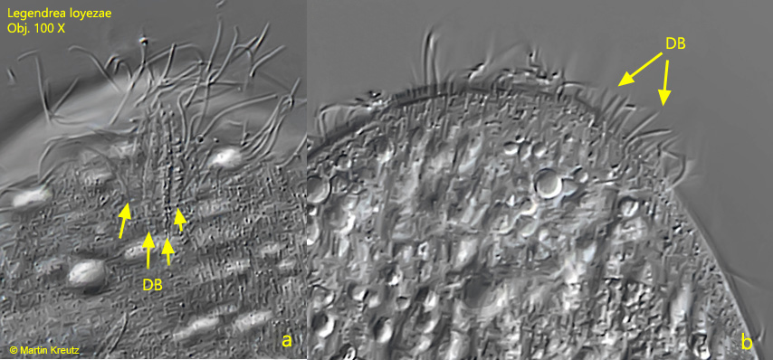
Unfortunately, in the cell I could not find any hint of any prey organisms or other food. Turning the specimen carefully under the coverslip I could see a four row dorsal brush with bristles about 5 – 6 µm long
Fig. 13 a-b:Legendrea loyezae. The dorsal brush (DB) in dorsal (a) and lateral view (b). Obj. 100 X.
Looking at the strange tentacles, one wonders what prey is caught with them and how the feeding actually takes place. An observation of a very large specimen (L = 120 µm without tentacles) at very high sample layer thickness gave a hint.
Fig. 14 a-b:Legendrea loyezae. The freely swimming specimen with a length of 120 µm shortly before entering the detritus flake. a) dorsal view, b) right lateral view. Obj. 60 X.
The specimen encountered a detritus flake under the coverslip and shortly thereafter it began to extend its tentacles. The tentacles started to explore a depression in the detrital flake, moving like snakes, apparently driven by the cilia at the distal end of the tentacles. The ciliate partially deformed to better reach the cavity. Unfortunately I could not observe the actual feeding process. So it remains unclear whether it is taken up directly by the tentacles (similar to a suctor) or is led from the tentacles to the mouth opening.
Recently a redescription of Legendrea loyezae was published by Weiss, Andreou and Esteban (s. Literature; Weiss, Andreou, Esteban 2022), in which they also document specimens with fully extended tentacles.
Fig. 15 a-d:Legendrea loyezae. a) the specimen is entering the detritus flake; b,c) the tentacles start to move like snakes to explore the cavity; d) the specimen is leaving the detritus flake with extended tentacles. Obj. 60 X.