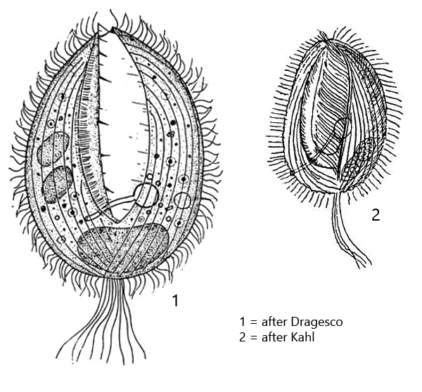
very large oral apparatus reaching down to posterior third
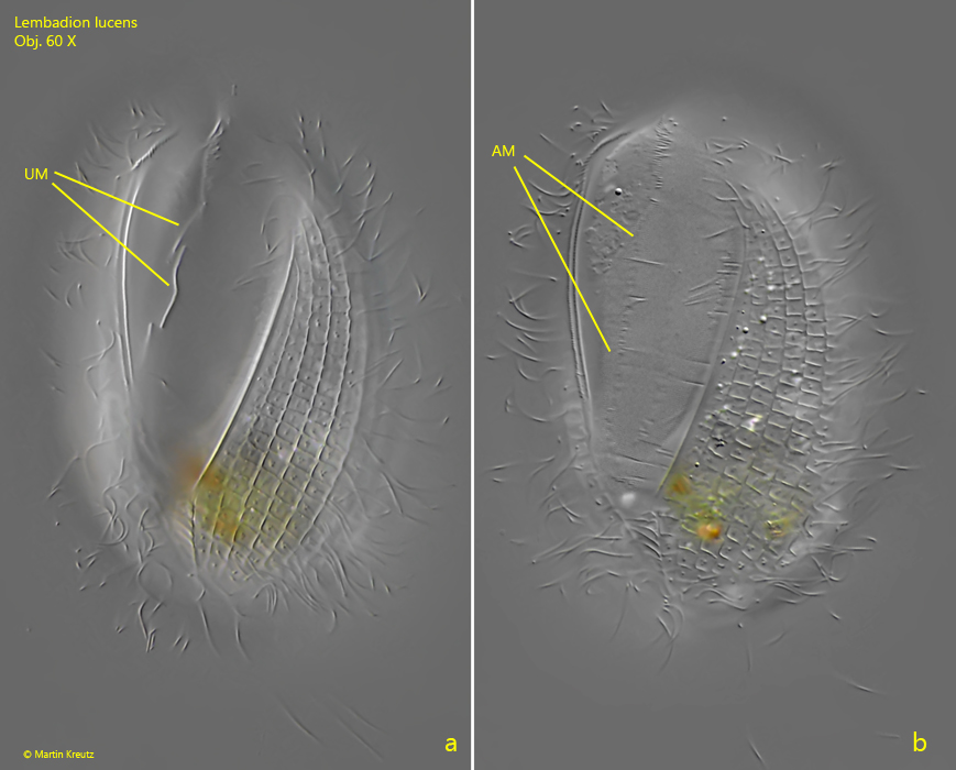
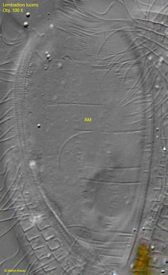
left side of oral apparatus an adorale membranelle of cilia glued together
right side of oral apparatus an undulating membrane (hard to see)
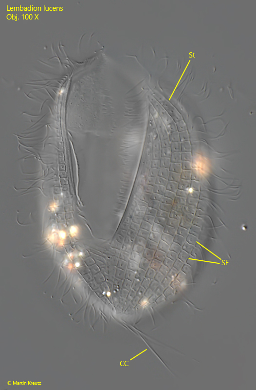
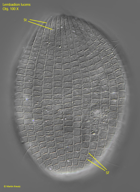
pellicle sculpture with pattern of regularly arranged squares, turns into stripes at the anterior end
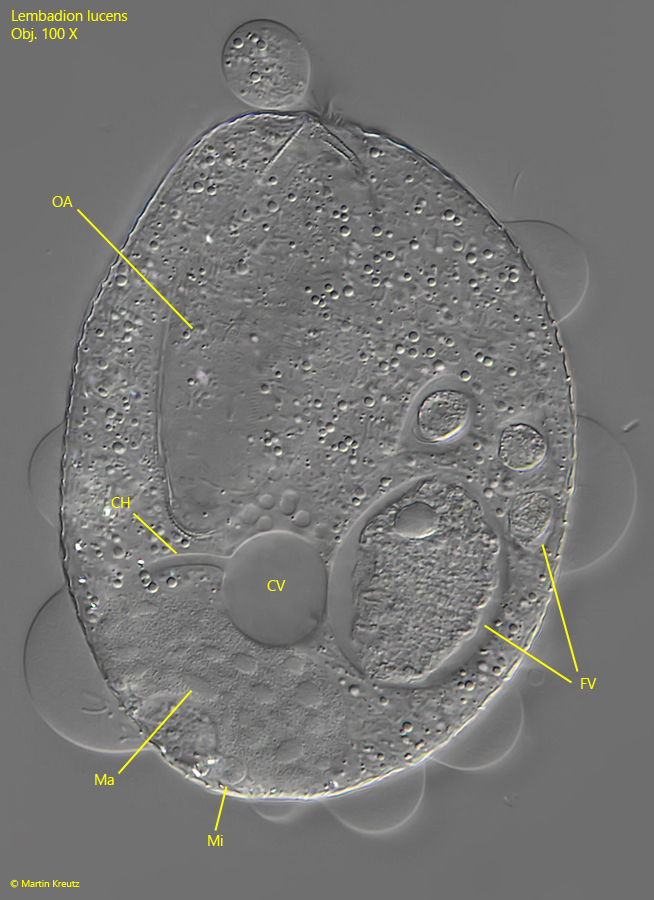
contractile vacuole is connected via a channel with the ventral excretion porus
macronucleus oval or kidney shaped in the posterior third with an adjacent spherical micronucleus
posterior end with a tuft of cilia, about 30 µm long
Lembadion lucens
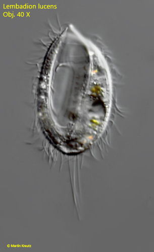
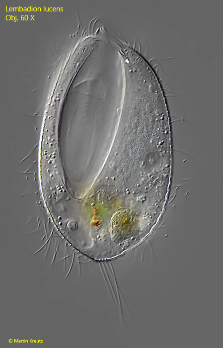
Lembadion lucens is a very common ciliate and is found in all my sampling sites. It is very easy to recognize by the large mouth opening, which reaches down to the posterior third of the body and the tuft of caudal cilia, which become quite long. Then at higher magnification you can see the pattern of regularly arranged squares in slightly squashed specimens. According to my observations this ciliate is not particularly fastidious with food and feeds on bacteria, algae and also small ciliates. This is in agreement with the observations of other authors. Lembadion lucens can be best distinguished from the similar species Lembadion bullinum by its size. If the length is smaller than 100 µm it is Lembadion lucens. The larger species Lembadion bullinum is 120 – 200 µm long. In my populations of Lembadion lucens the ciliate reaches an overall length of mostly 60 – 80 µm.
Fig. 1: Lembadion lucens. L = 57 µm. Ventral view of a freely swimming specimen. Obj. 40 X.
Fig. 2: Lembadion lucens. L = 80 µm. Ventral view of a second freely swimming specimen. Obj. 60 X.
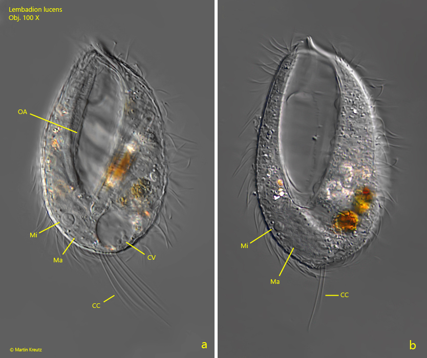
Fig. 3 a-b: Lembadion lucens. L = 60 µm. Ventral view of a slightly squashed specimen. CC = tuft of caudal cilia, CV = contractile vacuole, Ma = macronucleus, Mi = micronucleus, OA = oral apparatus. Obj. 100 X.
Fig. 4 a-b: Lembadion lucens. L = 80 µm. a) ventral view of a slightly squashed specimen with focus on the undulating membrane (UM). b) the same specimen with focus on the adorale membranelle (AM). Obj. 60 X.
Fig. 5: Lembadion lucens. The adoral membranelle (AM) consists of cilia glued together. These can only be visualized at highest magnification. They are arranged in an extremely regular manner. The interciliary distance is 0.38 µm. Obj. 100 X.
Fig. 6: Lembadion lucens. L = 60 µm. Ventral view of a squashed specimen with focus on the pattern of square fields (SF) of the pellicle. At the anterior end, the square fields turn into stripes (St). CC = caudal cilia. Obj. 100 X.
Fig. 7: Lembadion lucens. L = 60 µm. Dorsal view the same squashed specimen as in Fig. 6 with focus on the pattern of square fields (SF) of the pellicle. At the anterior end, the square fields turn into stripes (St). Obj. 100 X.
Fig. 8: Lembadion lucens. In this strongly squashed specimen the channel (CH) between the contractile vacuole (CV) and the excretion porus is visible. The excretion porus is located dorsally and not visible in this focal plane. FV = food vacuoles, Ma = macronucleus, Mi = micronucleus, OA = oral apparatus. Obj. 100 X.