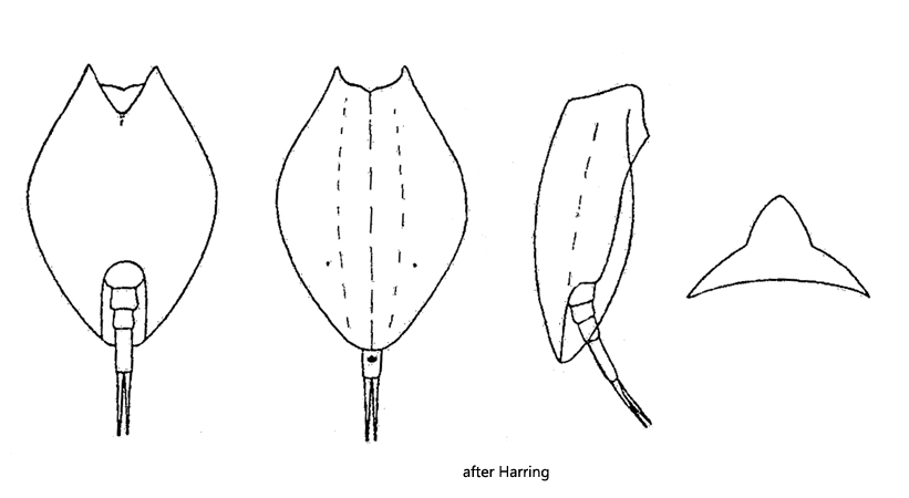
dorsally with a moderately high keel, sides of keel convex
ventral side flat with delicate longitudinal folds
ventral sinus V-shaped
foot groove U-shaped
three foot segments, terminal foot segment longer than basal section of foot
slender toes pointed
two eyespots
Lepadella rhomboides
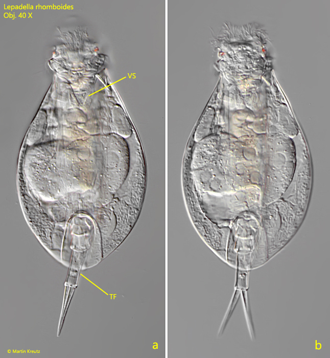
I have found Lepadella rhomboides so far exclusively in the Simmelried. However, the species is not very common. I documented findings in June 2004 and in July 2008. I recognize Lepadella rhomboides by the slender, oval shape, the V-shaped ventral sinus (s. fig. 1a) and the fine longitudinal folds on the ventral side of the lorica (s. fig. 3).
Fig. 1 a-b:Lepadella rhomboides. L = 120 µm (of the lorica). Ventral view of a slightly squashed specimen. Note the V-shaped sinus (VS) and the elongated terminal foot segment (TF). Obj. 40 X.
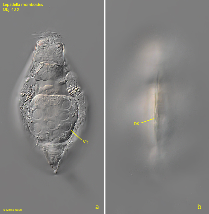
Fig. 2 a-b:Lepadella rhomboides. L = 115 µm (of the lorica). Two focal planes from dorsal. Note the ridge of the dorsal keel (DK). Vit = vitellarium. Obj. 40 X.
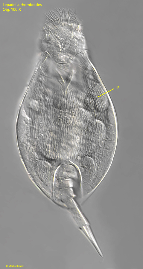
Fig. 3:Lepadella rhomboides. L = 85 µm (of the lorica). Ventral view of a slightly squashed specimen. Note the delicate longitudinal folds of the lorical (LF). Obj. 100 X.
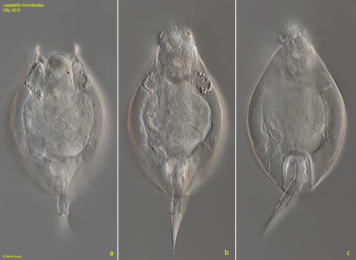
Fig. 4 a-c:Lepadella rhomboides. L = 127 µm (of the lorica). Ventral view of a slightly squashed specimen found in November 2022. Obj. 60 X.
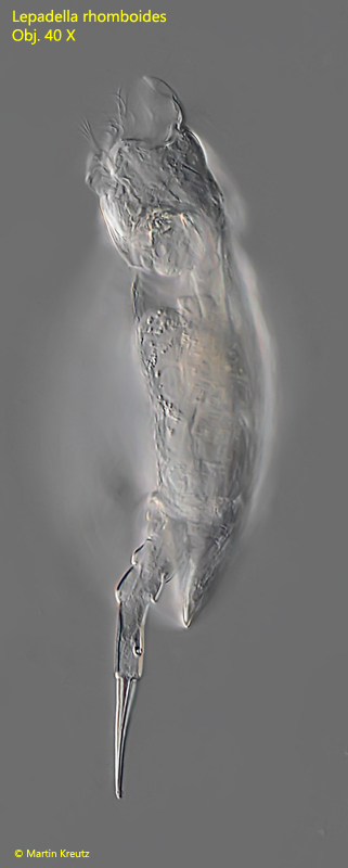
Fig. 5:Lepadella rhomboides. L = 115 µm (of the lorica). Lateral view from left of a freely swimming specimen. Obj. 40 X.