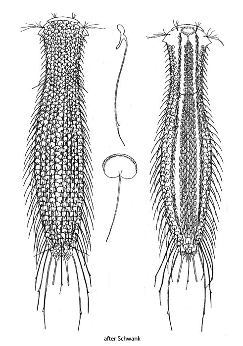
cephalion present two pairs of dorsal setolae, posterior ones on special oval scales
toes 30–39 µm long with thin adhesive tubes (20–33 µm)
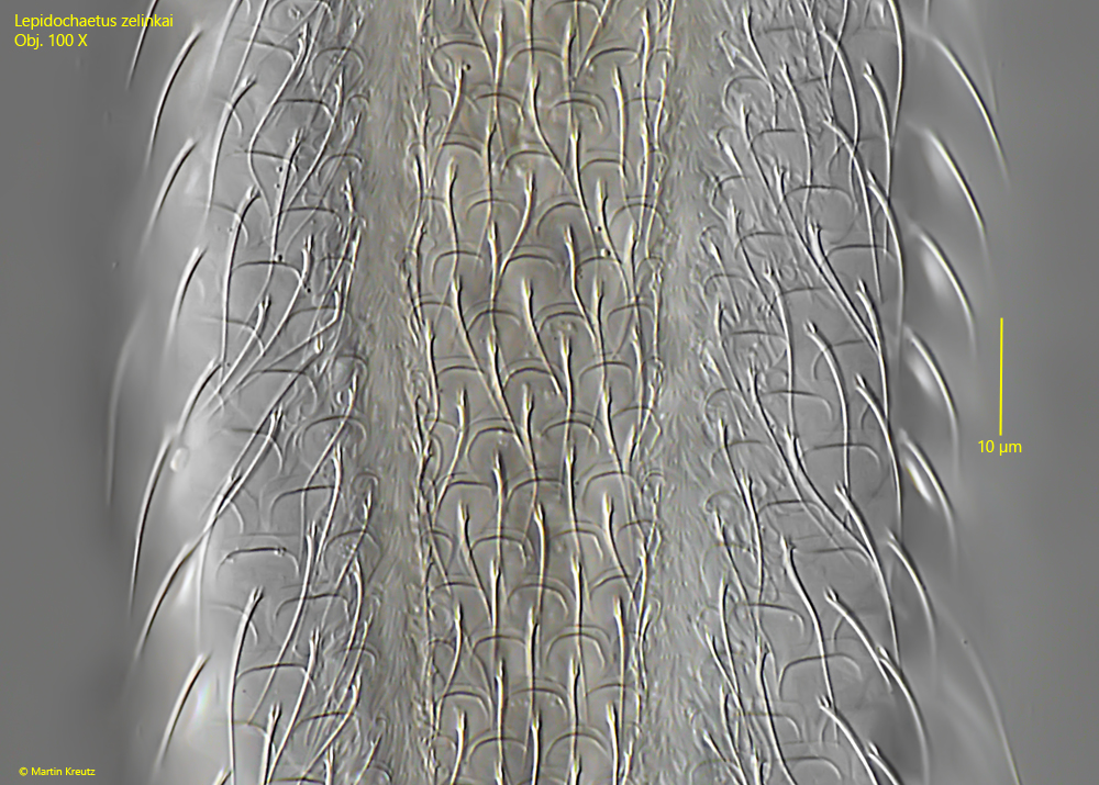
dorsal 11–13 longitudinal rows of oval or elliptically scales with a bulged anterior margin
dorsal scales with simple spines, 30–35 µm long
ventral 7 longitudinal rows of oval scales with short, simple spines
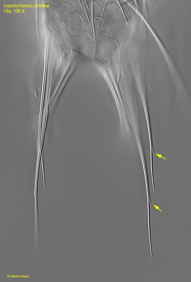
at posterior end 7 elongated spines each with a secondary spine, 30–85 µm long
dorsal anal region covered with roundish, spineless scales
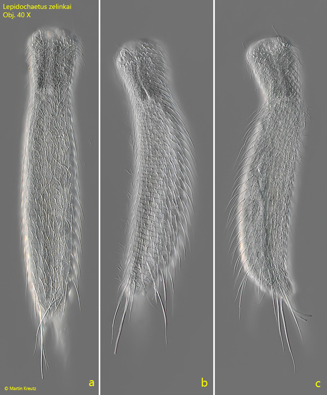
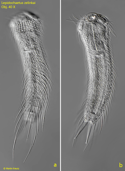
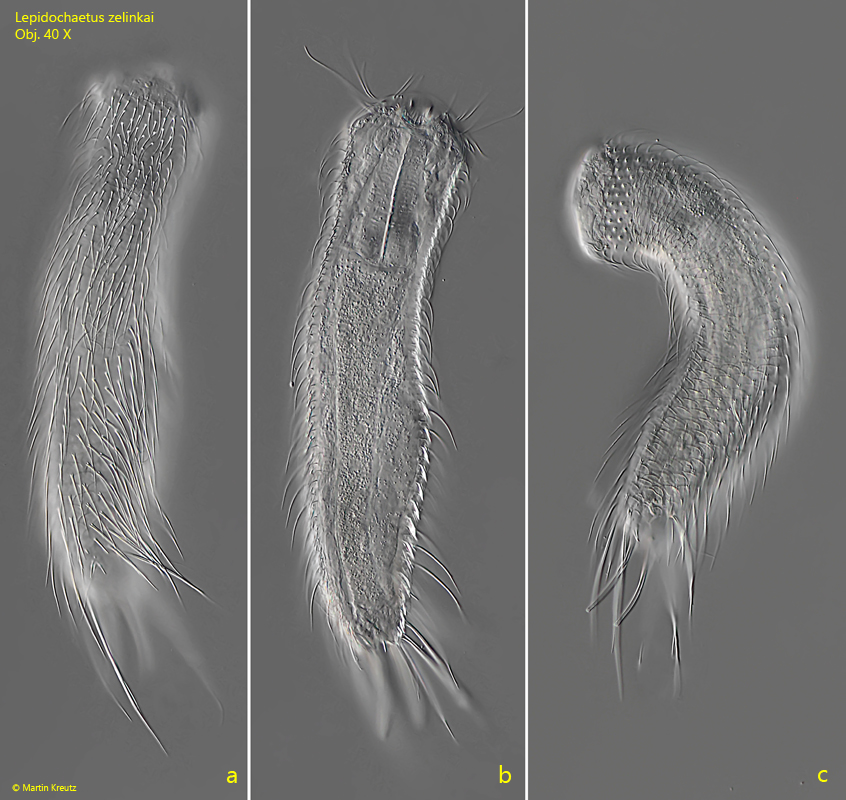
Lepidochaetus zelinkai
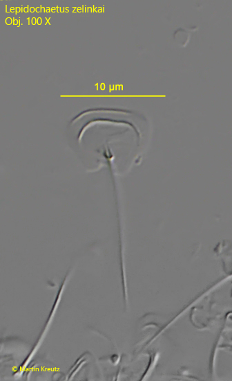
So far I have only found Lepidochaetus zelinkai in the uppermost mud layer in the Simmelried. Lepidochaetus zelinkai can be recognized even at low magnification by the 7 elongated terminal spines (s. fig. 9). Three pairs arise laterally and one spine arises dorsally in the middle of the anal region. The dorsal scales have a raised anterior margin and a distal indentation, which can only be recognized on isolated scales (s. fig. 6). The ventral scales are smaller versions of the dorsal scales (s. figs. 7 a-b and 8). However, the simple spines are shorter here.
Fig. 1 a-c:Lepidochaetus zelinkai. L = 204 µm (without terminal spines). A freely swimming specimen from dorsal (a, b) and from right (c). Obj. 40 X.
Fig. 2 a-b:Lepidochaetus zelinkai. L = 215 µm (without terminal spines). A second freely swimming specimen from dorsal. Obj. 40 X.
Fig. 3 a-c:Lepidochaetus zelinkai. L = 238 µm (without terminal spines). A third freely swimming specimen from dorsal. Obj. 40 X.
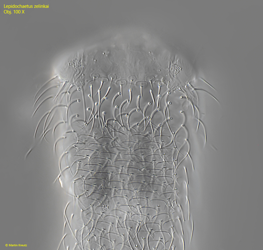
Fig. 4:Lepidochaetus zelinkai. The dorsal scales covering the head in detail. Obj. 100 X.
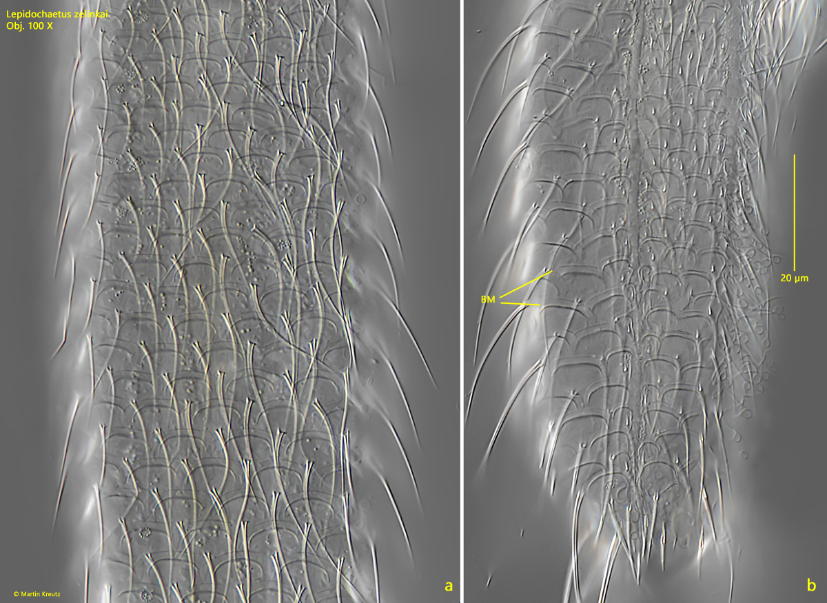
Fig. 5 a-b:Lepidochaetus zelinkai. The dorsal scales of a slightly squashed (a) and squashed specimen (b). Note the bulged anterior margin of the scales (BM). Obj. 100 X.
Fig. 6:Lepidochaetus zelinkai. A detached dorsal scale. Obj. 100 X.
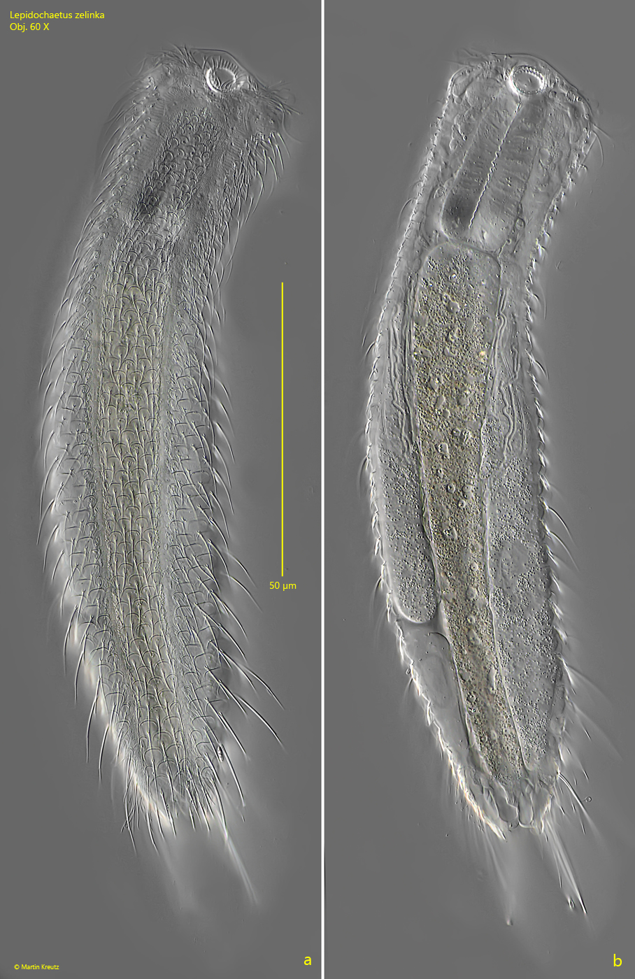
Fig. 7 a-b:Lepidochaetus zelinkai. L = 304 µm (without terminal spines). Two focal planes from ventral. Obj. 100 X.
Fig. 8:Lepidochaetus zelinkai. The ventral scales in detail. Obj. 100 X.
Fig. 9:Lepidochaetus zelinkai. The elongated, terminal spines, each with a small secondary spine (arrows). Obj. 100 X.