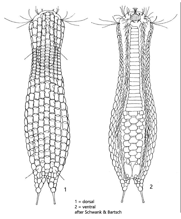
two pairs of dorsal setolae, posterior one on special scales, sometimes double-keeled.
toes completely scaled, 18–27 µm long
adhesive tubes half of toe-length
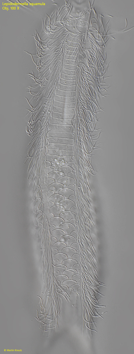
dorsally 7–9 longitudinal rows of spineless scales, maximal size 7– 10 X 4–10 µm
proximal margin of scales strongly convex and overlapping
head and trunk scales elongately oval
neck scales transversely oval and very short (4–5 x 8–9 µm)
at posterior end 2 large terminal scales and numerous small scales
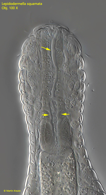
pharynx cylindrical, terminally only slightly swollen, 28-56 µm long
Lepidodermella squamata
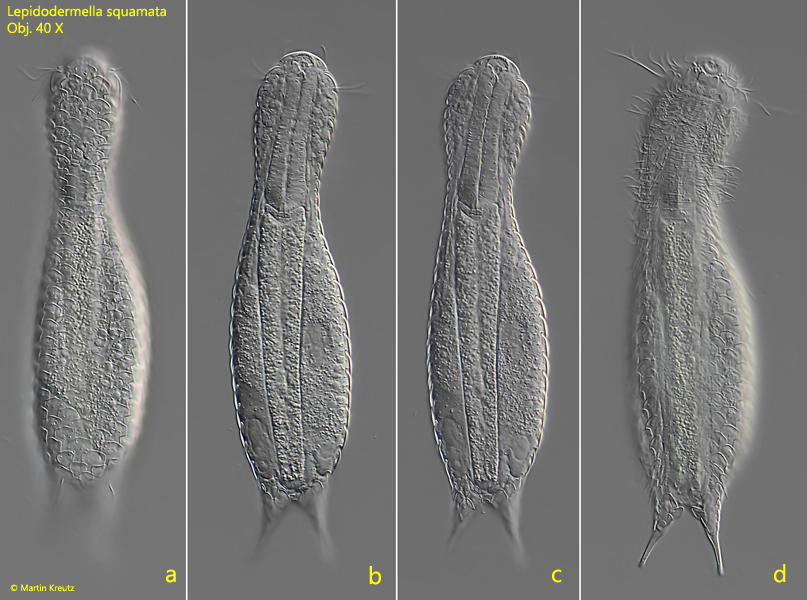
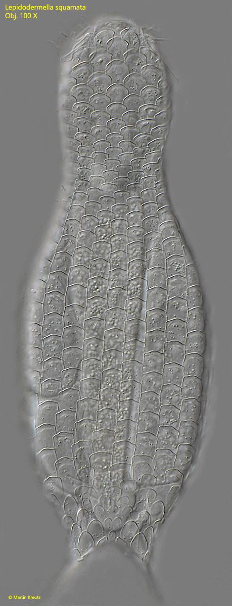
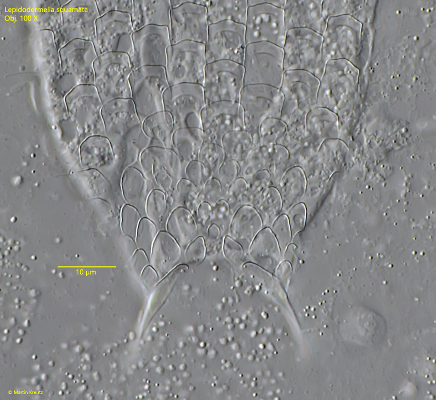
Lepidodermella squamata is supposed to be one of the most common gastrotrichs. However, I only found one specimen in August 2022 in rotting plant masses from Simmelried. The species is easily recognized by the dorsal scales, which are arranged like shigles on a roof and overlap strongly. They do not possess a keel or a spine. In addition, the toes are also covered with scales. On the ventral side, the scales in the anterior third are arranged in broad bands, which is also very characteristic. On the dorsal surface of the pharynx I could recognize a structure reminiscent of a tuning fork, which is not described yet for Lepidodermella squamata. However, it has been observed in other gastrotrichs (e.g. Chaetonotus hoanicus, Müller, 2022).
Fig. 1 a-d:Lepidodermella squamata. L = 185 µm. Dorsal view of a freely swimming specimen. Obj. 40 X.
Fig. 2:Lepidodermella squamata. L = 185 µm. Dorsal view of squashed specimen. Obj. 100 X.
Fig. 3:Lepidodermella squamata. L = 185 µm. The dorsal scales at the posterior end in detail. Obj. 100 X.
Fig. 4:Lepidodermella squamata. L = 156 µm. The ventral scales of a slightly squashed specimen. Obj. 100 X.
Fig. 5:Lepidodermella squamata. L = 185 µm. Peculiar tubes reminiscent of a tuning fork with unknown function are visible on the dorsal surface of the pharynx visible (arrows). Obj. 100 X.